Tag Archives: time-lapse

Watching Bacteria Grow: Winogradsky Panel Day 49

Various green bacteria are growing now all over the panel.  These are likely to be cyanobacteria and some green sulfur bacteria.

IMG_6151

You may also notice the addition of clamps along the bottom.  On Day 33, I arrived to find a puddle below the panel and all of the water drained out.  Luckily, the crack was too small to let any soil out.  The clamps are holding well so no more leaked out after I refilled it.  Perhaps I can claim that this is an experiment in shocking the ecosystem with a drying and aeration event?  I didn’t notice any significant changes in the following days and weeks, except the continued growth of green and yellow bacteria, so I suspect the shock wasn’t too detrimental.

IMG_6156

In the last several days, a filamentous green colony has developed.  Note the large gas bubbles around it.  This is likely a cyanobacteria like Oscillatoria, so the gas would be oxygen generated through its plant-like photosynthesis.  IMG_6120IMG_6141The two images above show the top right corner of the panel at day 35 and then day 49.  Notice how the black mud is slowly being covered by the growth of a very diverse mixture of microbes.

Share

Watching Bacteria Grow: Winogradsky Panel Day 30

The pace of the changes in the Winogradsky panel has slowed, but now some green microbes are really noticeable.  There are shades of bright blue-green, yellow-greens and yellow.IMG_6117IMG_6118IMG_6120IMG_6121

The range of greens is due to different combinations of pigments in photosynthetic bacteria.  I had a recent conversation with someone who didn’t realize that organisms other than plants can photosynthesize.  Many bacteria can, and like plants, they use a variety of different pigments to capture energy from light and use it to drive metabolic reactions in the cell.  The green is from chlorophylls, the same as plants use.  These are found in the cyanobacteria, which have the deeper blue-green color.  But there are many others too:  the “purple bacteria” have bacteriocholorophills and carotinoids, giving them a purple, yellow or brown color, and the bacteriochlorophylls of the “green sulfur bacteria” produce green or brown colors.  The role of these pigments is to absorb different wavelengths of light to harvest the energy.

Phototrophy in cyanobacteria is most similar to that of plants.  In fact, the chloroplast, the organelle found in plant cells that carries out photosynthesis, is very similar to cyanobacteria.  Most likely, early in the evolution of life, a larger cell engulfed an ancestral cyanobacterium and a symbiotic relationship evolved, eventually becoming plants.  Like plants, cyanobacteria use water in the process of capturing light energy, producing oxygen.  Some of the gas pockets seen in the Winogradsky might be oxygen.  Other bacteria use a slightly different process, and use hydrogen sulfide instead of water, and produce sulfur.  The sulfur forms globules, which are stored inside or outside the cell.

Not all color is from pigmentation of cells however.  The red-oranges in the Winogradsky panel are likely due to a combination of pigmented cells but also due to the oxidation of iron.  Some bacteria, like Chromatium, are able to use iron in phototrophy, instead of hydrogen sulfide or water.  They oxidize the iron in the soil, and oxidized iron has a red-orange color (the rust on a car is oxidized iron).


Share

Watching Bacteria Grow: Winogradsky Panel Day 14

There are many interesting changes over the last few days. The black areas in the sulfur enriched mud are coalescing, and there are some light pinkish-red colonies developing at the surface. The olive-green band next to the orange is either disappearing or getting covered up as the orange stripe expands.

IMG_6013

The unenriched mud is developing more visible colonies too, as various browns and some very pale greens start to appear.  A few emerald greens are now visible, most of them very faint but there is one larger bright one with a big gas bubble.IMG_6014
It turns out that the bloodworms (midge larvae) are not dead.  In the past, when I have prepared columns (instead of a panel), they all die within the first few days.  Here, they seem to be doing quite well.  Notice the worm tracks in the top left (photos taken Day 11).  It will be interesting to see how the bacterial colonies develop in this area in the presence of this disturbance.

IMG_5999

Share

Watching Bacteria Grow: Winogradsky Panel Day 7

The panel is changing more slowly now, but the oranges and greens are getting richer and the sulfur-enriched mud is showing more and coalescing black spots.
IMG_5962The level of the mud dropped overnight.  This could be due to settling of the mud and/or release of gas bubbles.  There are some water-filled gaps, and the water level is lower.  There is now air above the entire length of the water surface, instead of just at the opening of the panel (top left).  Note that there is also a left to right gradient in the water, which was already present before settling.  Near the opening, there is brownish/red colonies growing on the walls, probably aerobes (requires oxygen) explaining why its not present further from the opening.  The water is murky from left to right, perhaps microbes that can tolerate low levels of oxygen, up to a point near the right side where the water layer is still clear.

IMG_5965
IMG_5967
IMG_5968


Share

Watching Bacteria Grow: Winogradsky Panel Day 4

The Winogradsky panel is developing much more quickly than I had expected. Differences are noticeable from day to day.

IMG_5928
The panel at day 4.

IMG_5933
The colors at the interface are richer and thicker. The green sulfur bacteria below the orange layer are more obvious now.

IMG_5937The modified mud layer now has more black spots and there are now some bright green spots, probably green sulfur bacteria.  GSB are more tolerant of high H2S concentrations than the purple sulfur bacteria, so are growing well in the areas where the black spots tell us there is H2S.  Note the gas bubbles at the bottom center of the image.  The squiggly lines are actually paths left behind by the movement of small, red larvae of midge flies, called blood worms.  They made their way up to the top, and are now all apparently dead.  (And will be food for some bacteria).

IMG_5944


Share

Watching Bacteria Grow: Winogradsky Panel

One of the research projects in my lab is to investigate the microbial communities present in Winogradsky columns. You can read more about what a Winogradsky column is here.  Over the next little while, I will be carefully watching the development of the microbial communities over time.  This will help me set up a future experiment in which I will take samples over time and determine what bacteria are present and at what abundance to see how the community changes.  I will be posting regular updates as the Winogradsky panel develops.

IMG_5882
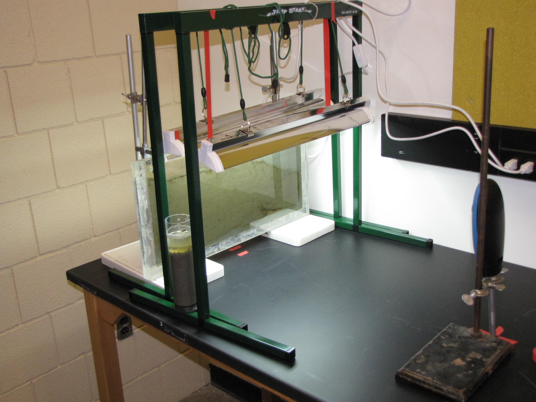
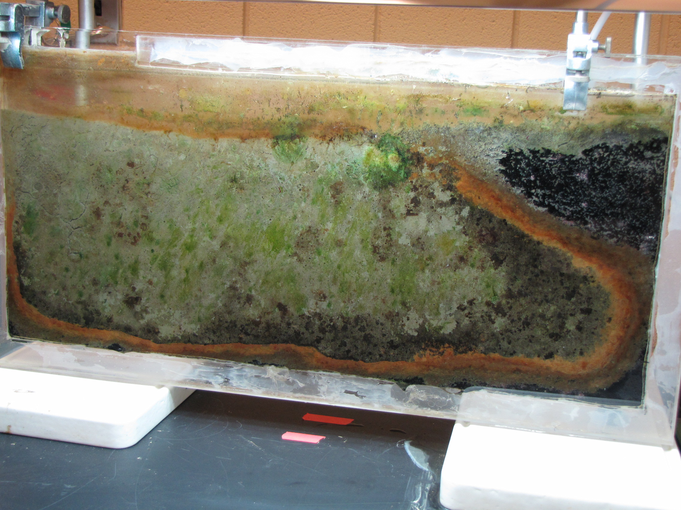
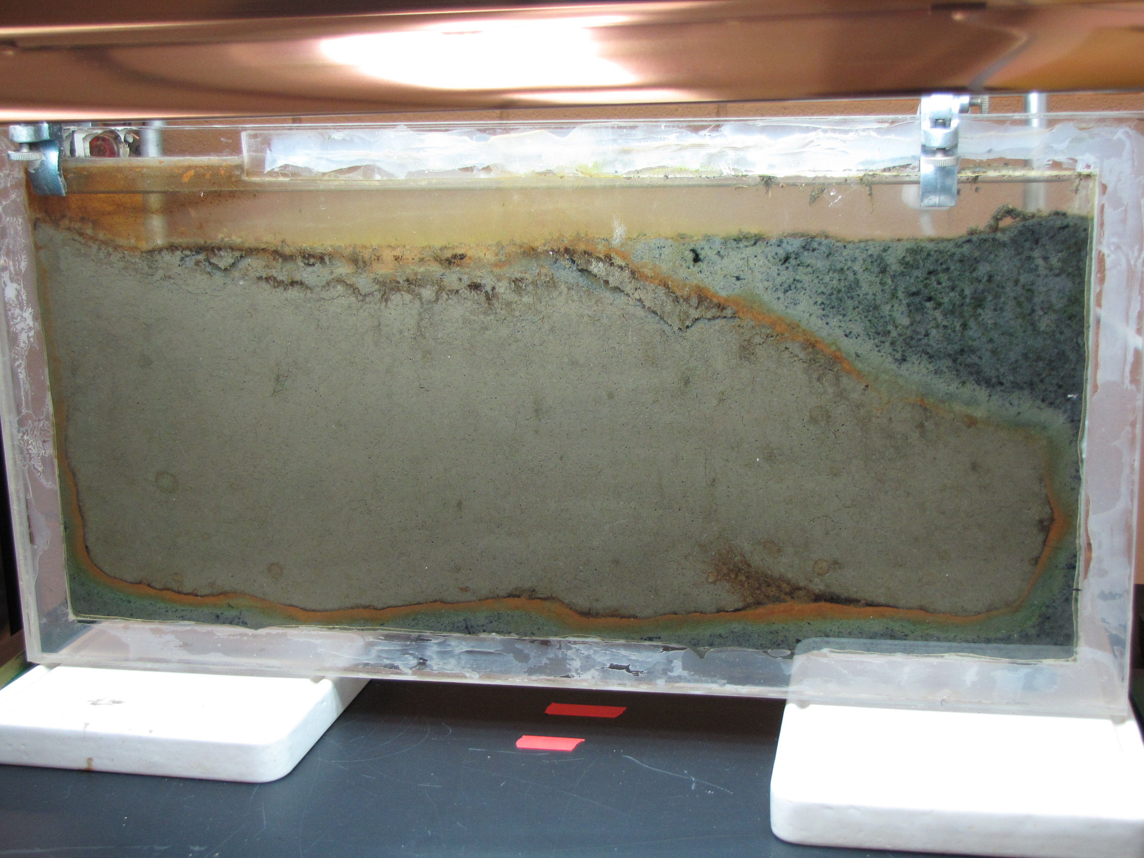
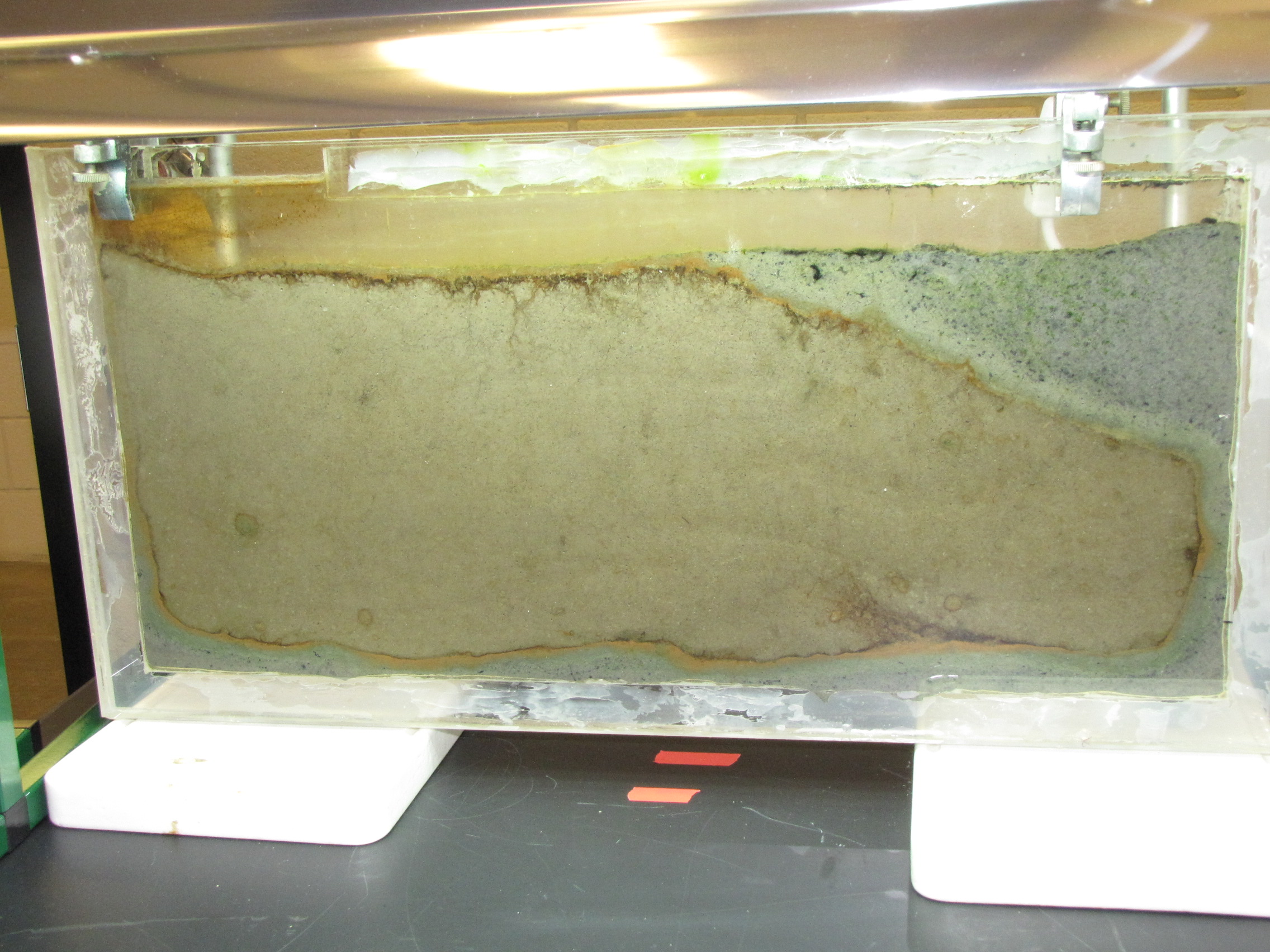
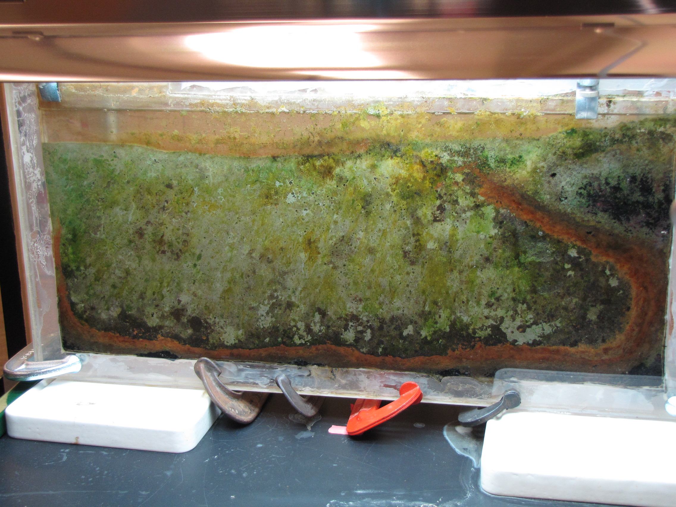
The set-up: the panel is 1′ x 2′ plexiglass with a 1/4″ gap filled with mud from a pond on Vassar campus, grow lamps, and time-lapse camera.

IMG_5883
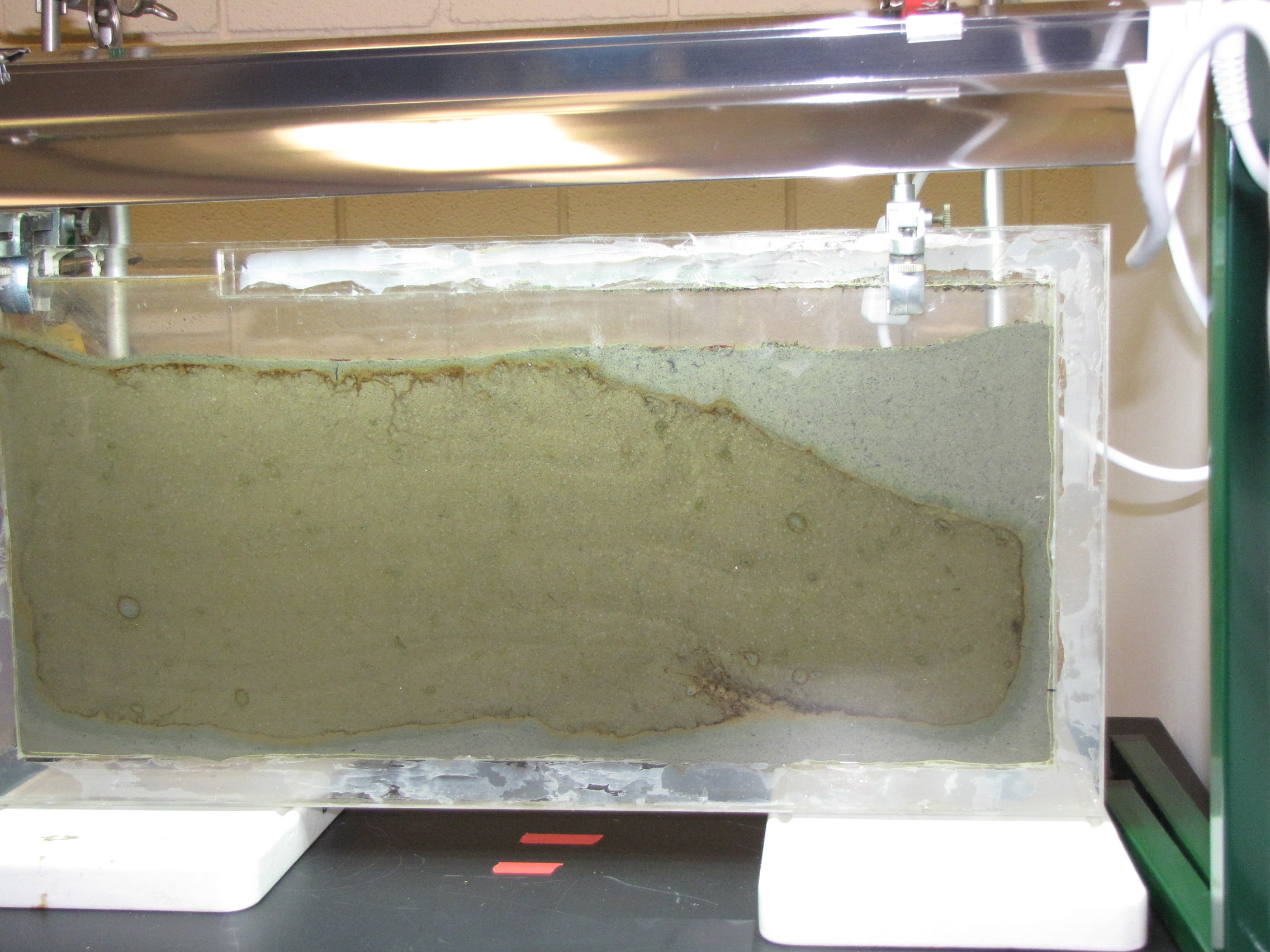
The panel after 2 days. The panel was prepared by adding calcium carbonate, calcium sulfate, and dried leaves to mud from a pond on Vassar campus.  We sifted the mud to make a fine slurry.  We collected leaves from near the pond and baked them to dry them, then blended in a blender to make as fine a powder as possible.  The leaves serve as a source of cellulose.  This supplemented mud was poured to a depth of about 4 cm.  Then we poured unmodified mud (no additives) on top.  Instead of just layering on top though, the bottom layer was displaced, and most of it was pushed to the right side and up to the top.  You can see it clearly here: the mud with additives has the lighter grey color.

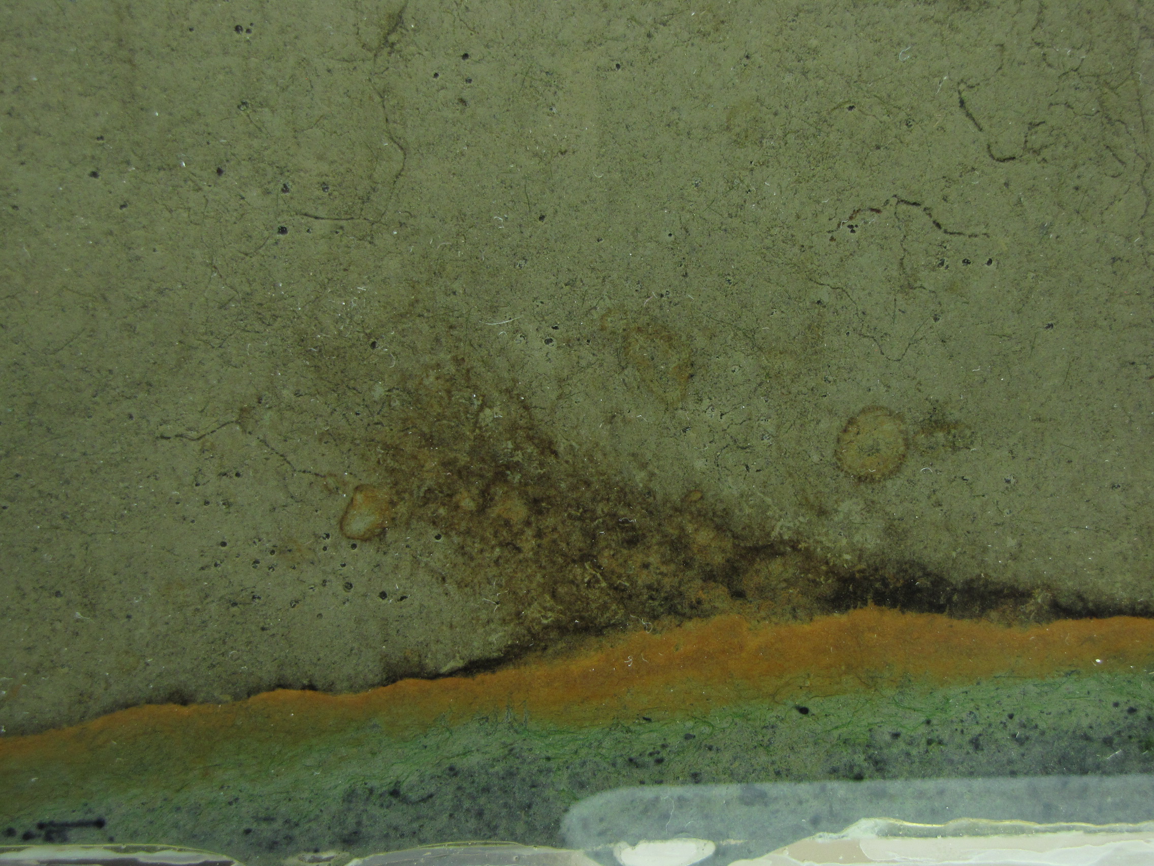
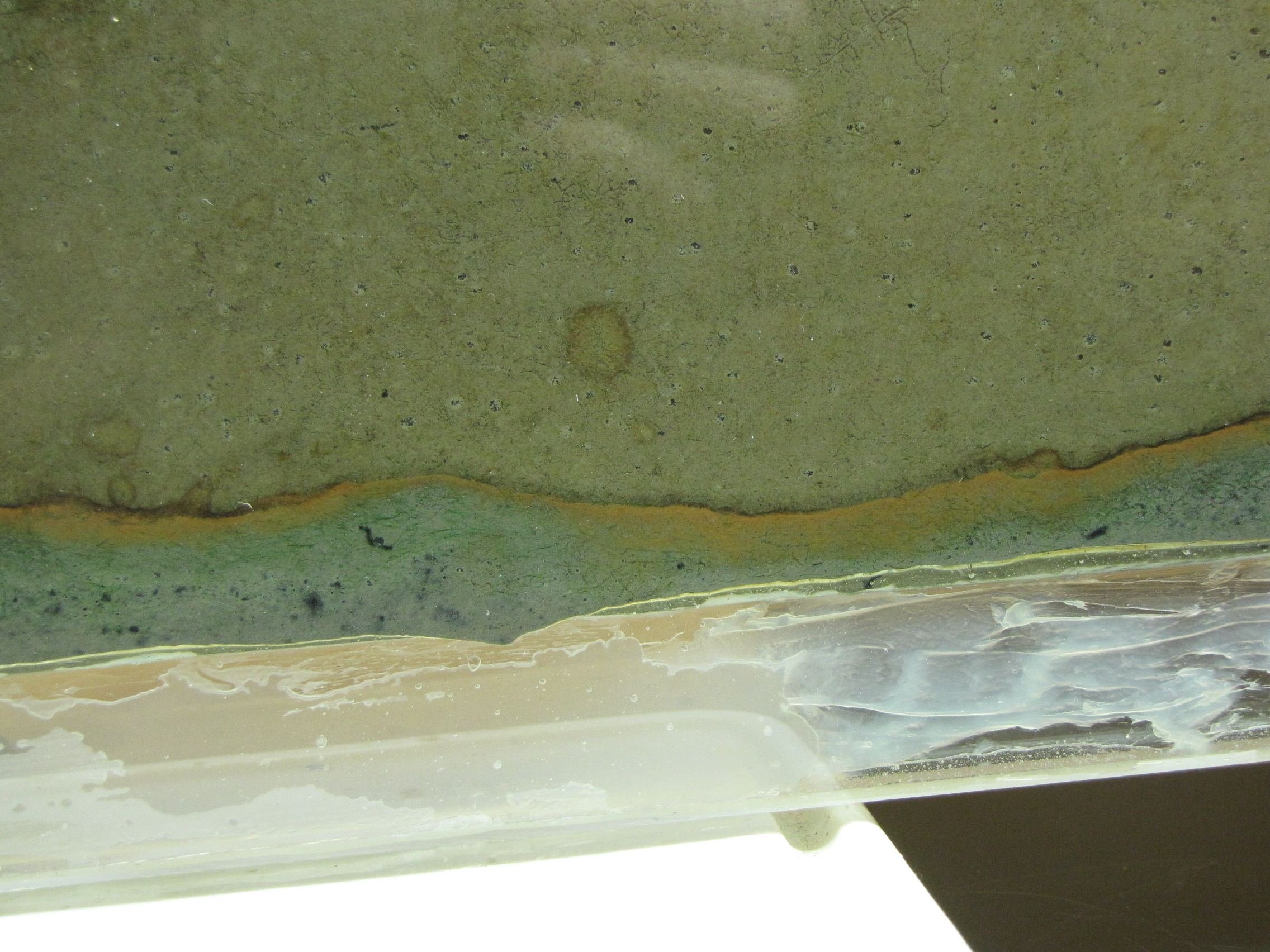
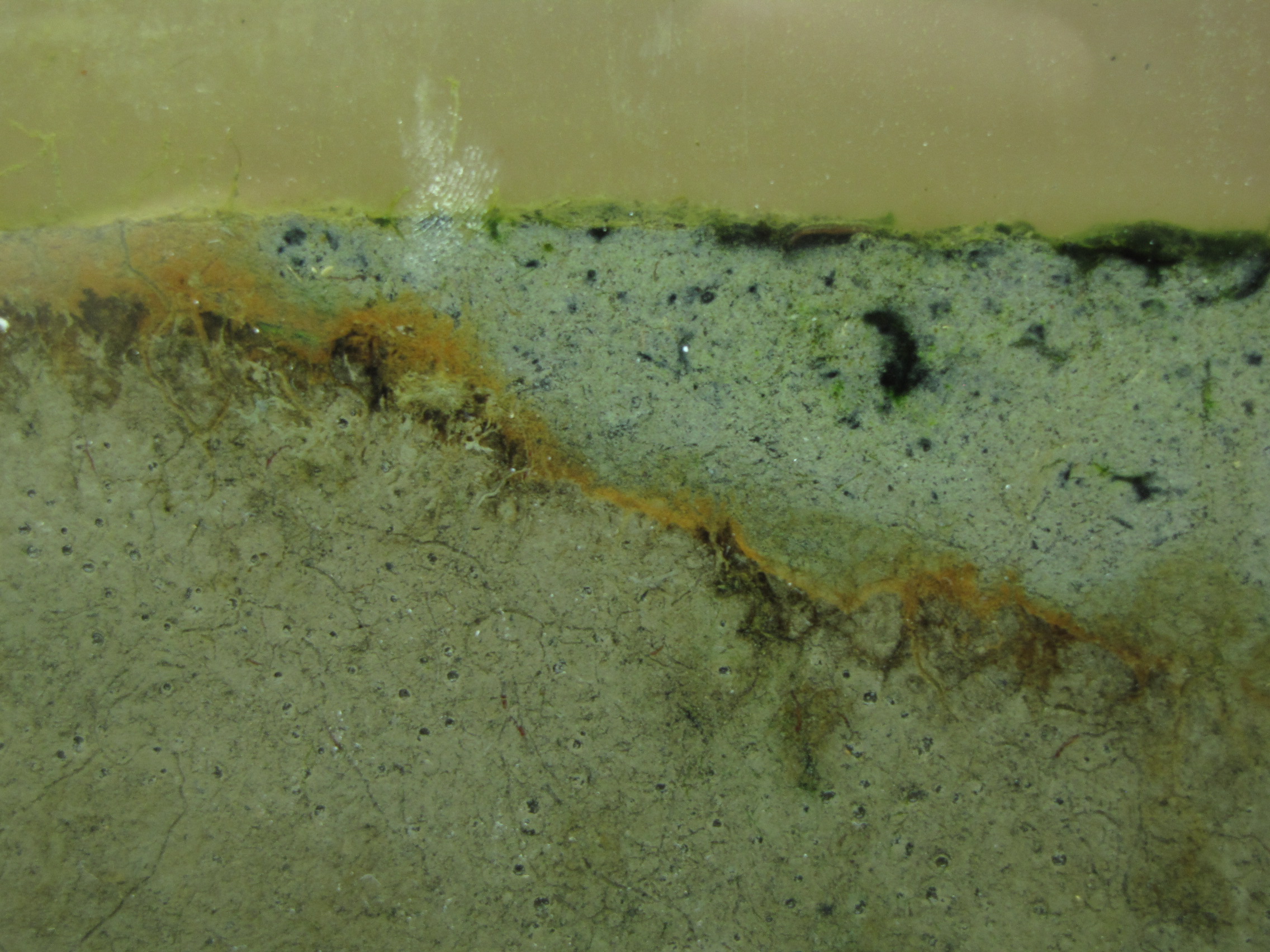
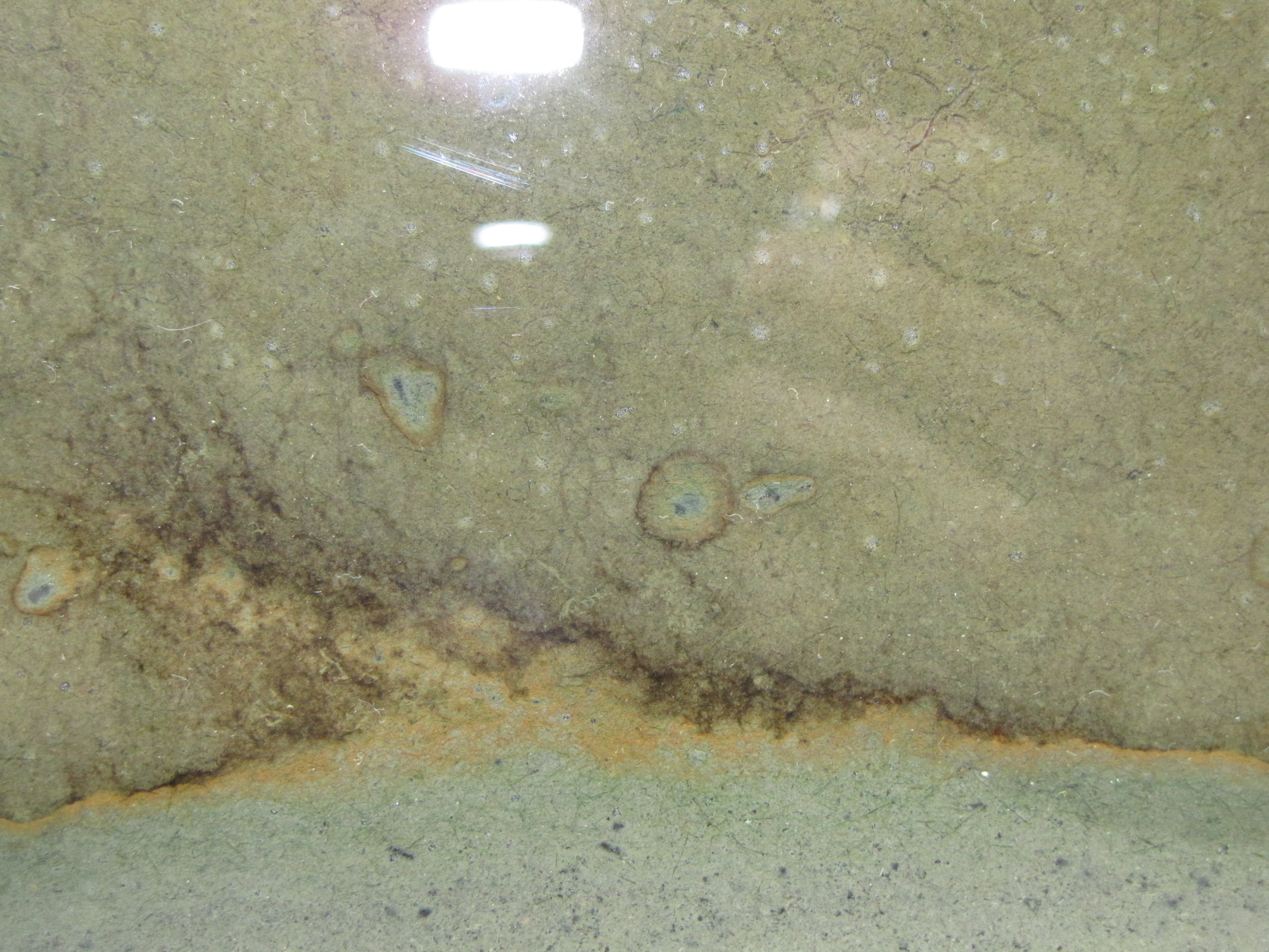
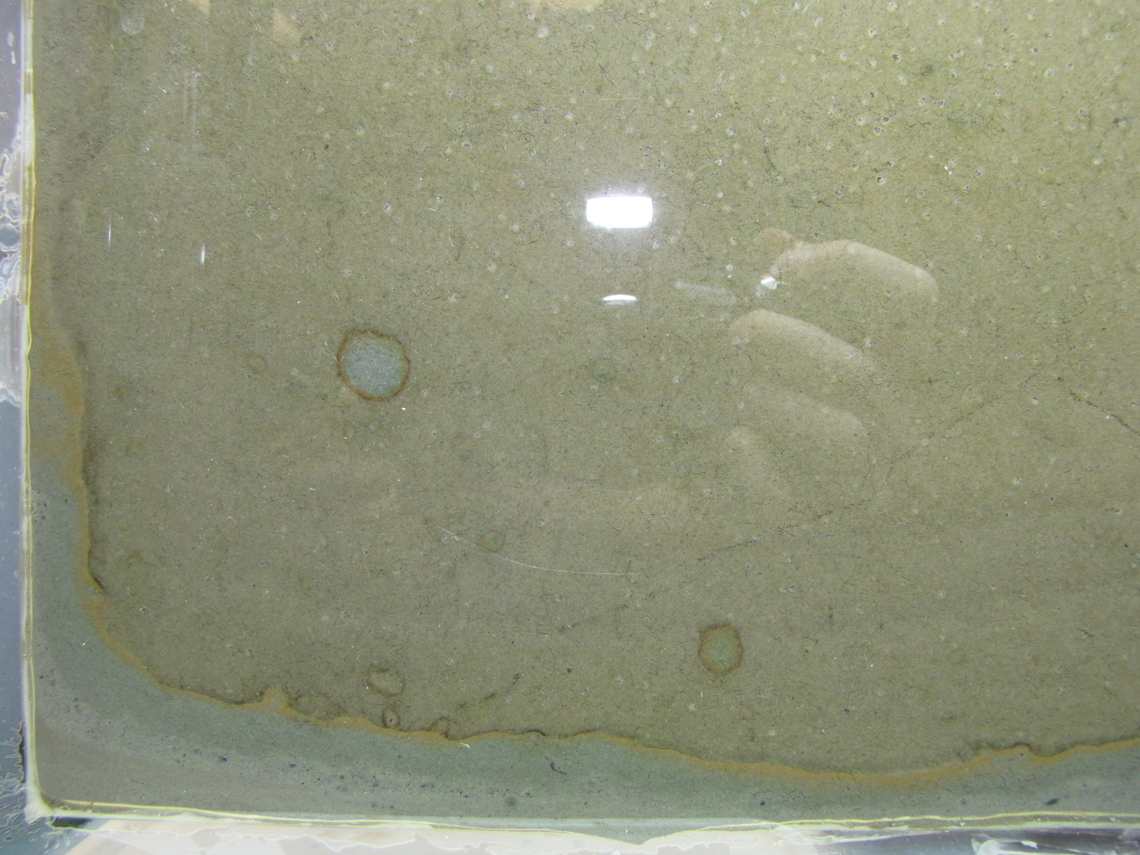
Note the orange and brownish-orange along the junction between the two muds. This is microbial growth, probably the phototrophic “purple bacteria.” Purple bacteria come in many colors, including red, orange, brown, and yes, purple.  They use light for energy but unlike plants they use H2S instead of water (H2O) as an electron source, and produce sulfur instead of oxygen.  You can also see it in the little circles where a spot of sulfur-enriched mud got surrounded by unmodified mud.

IMG_5886
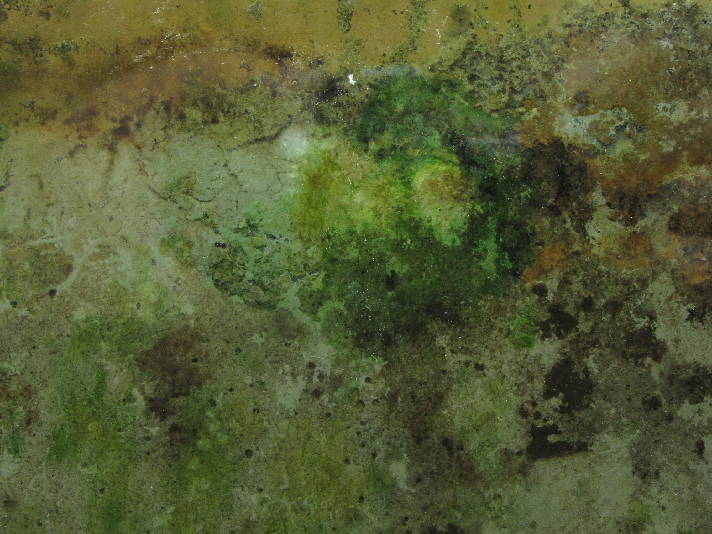
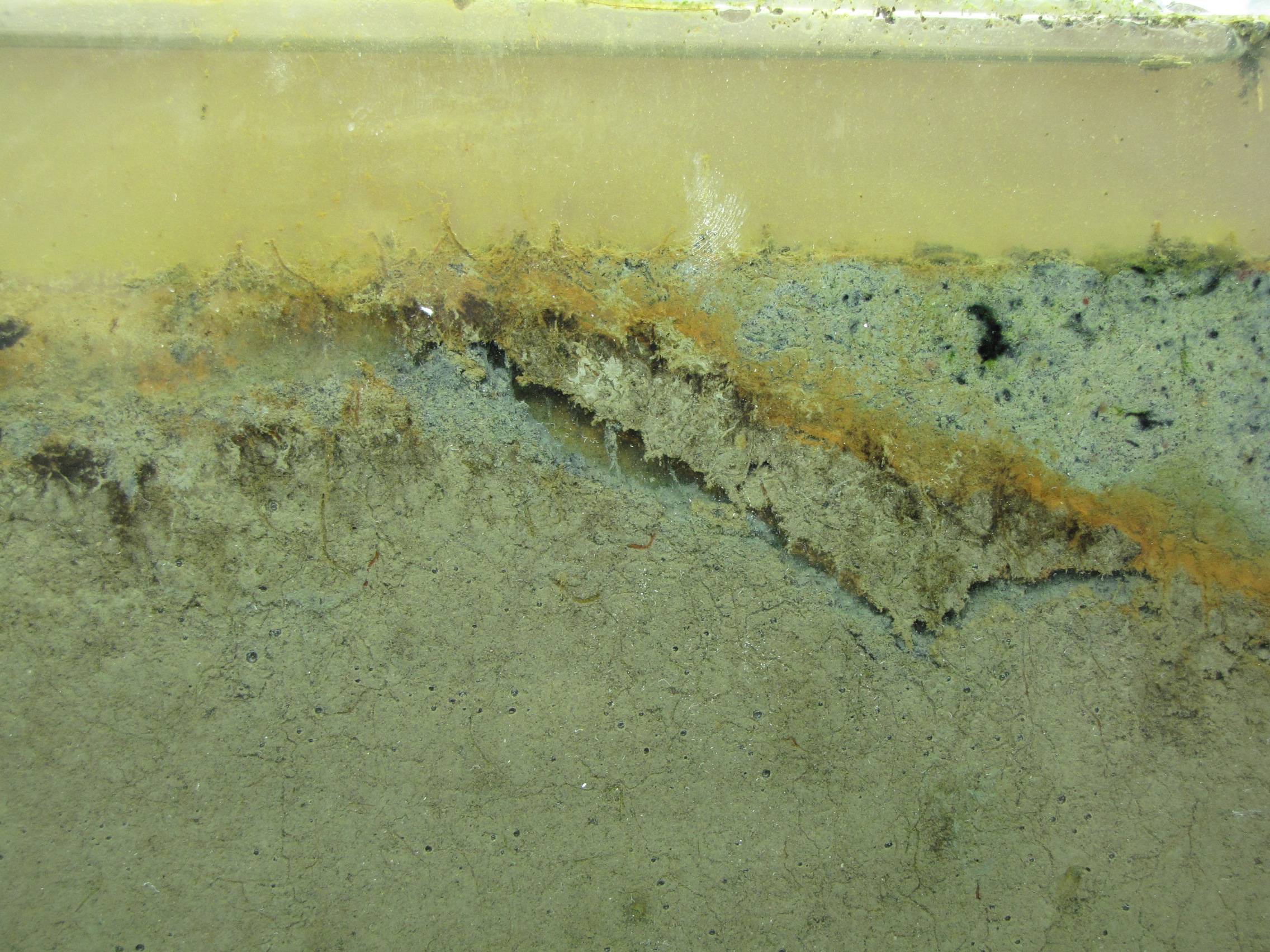
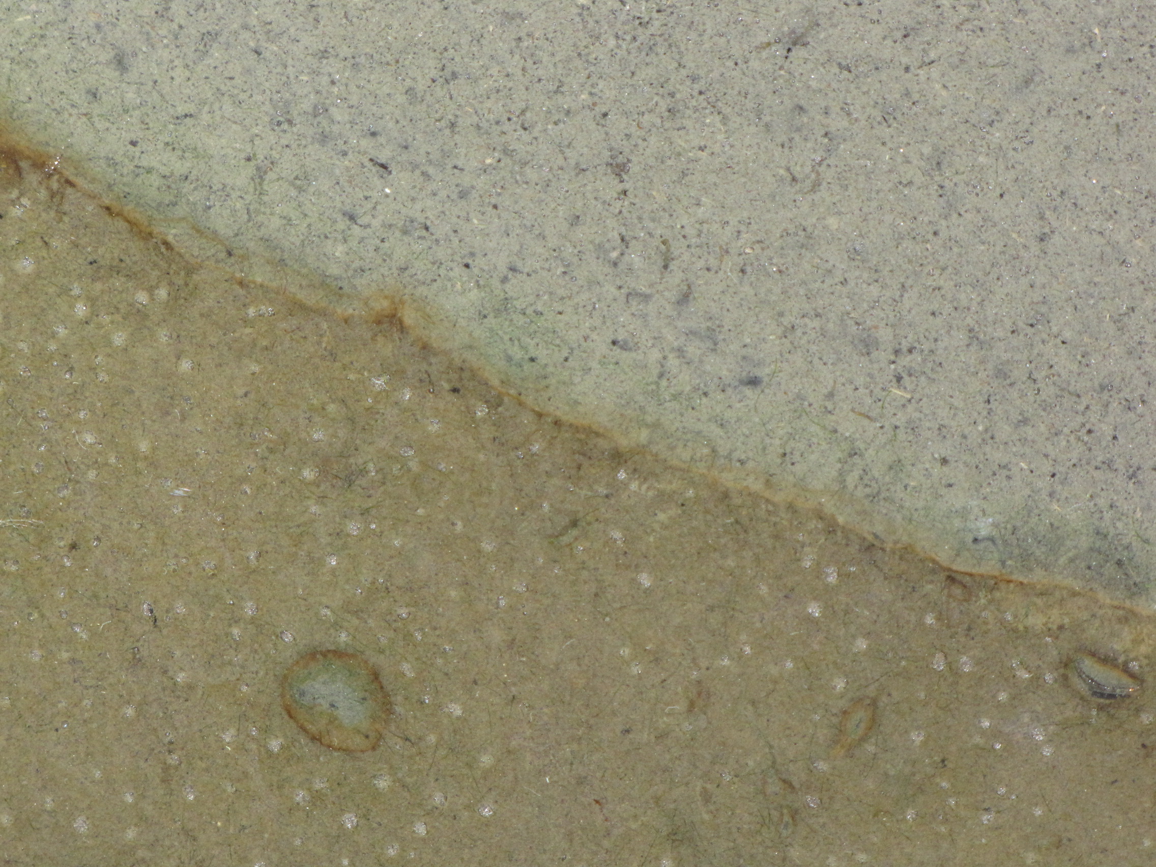
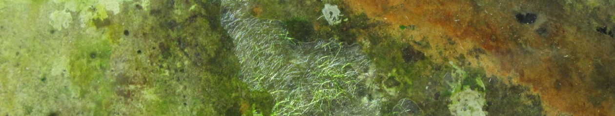
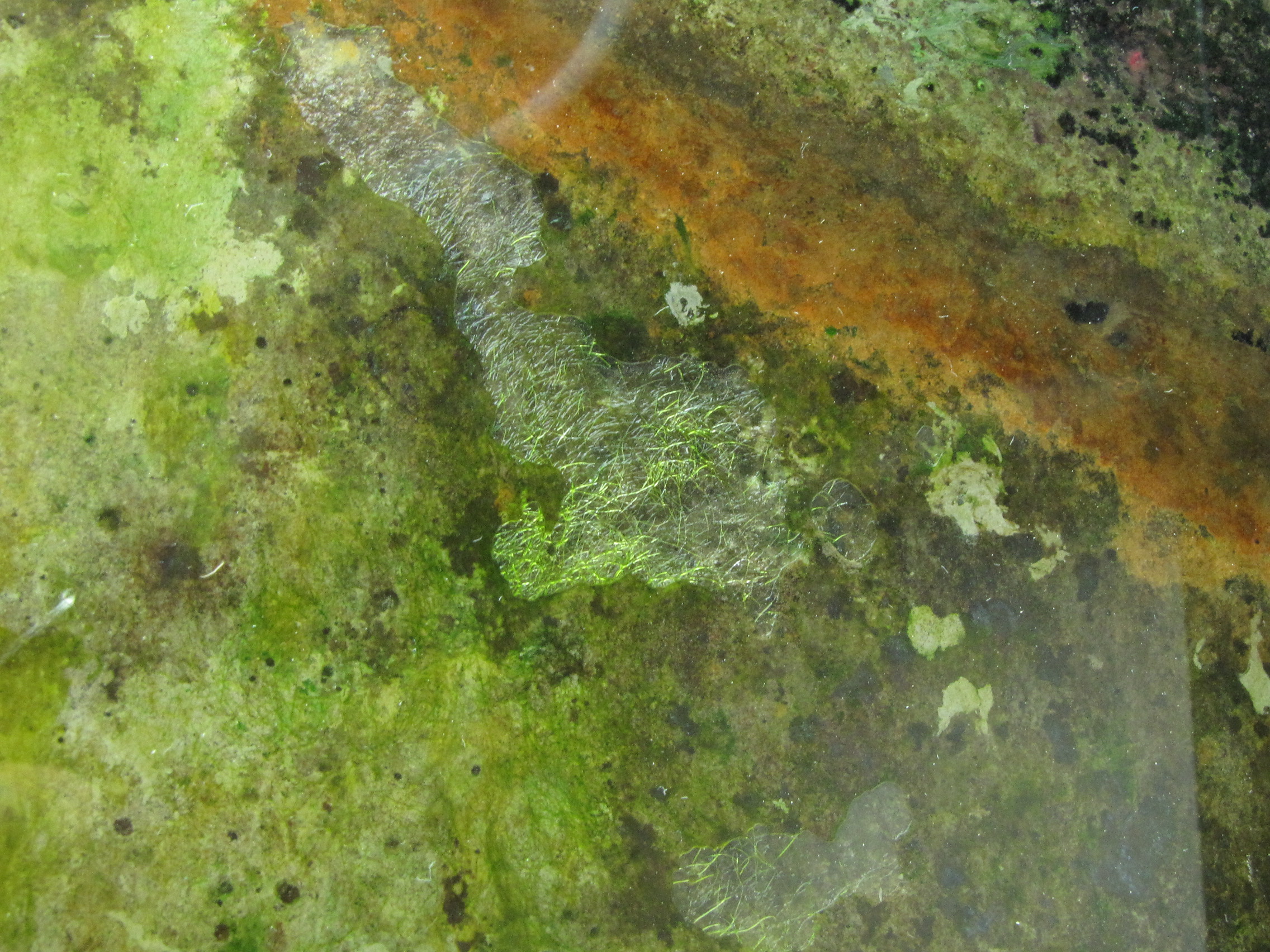
Close-up of the layers. There is a faint hint of green below the orange layer, these are green sulfur bacteria.

IMG_5898
Another close-up.

IMG_5884
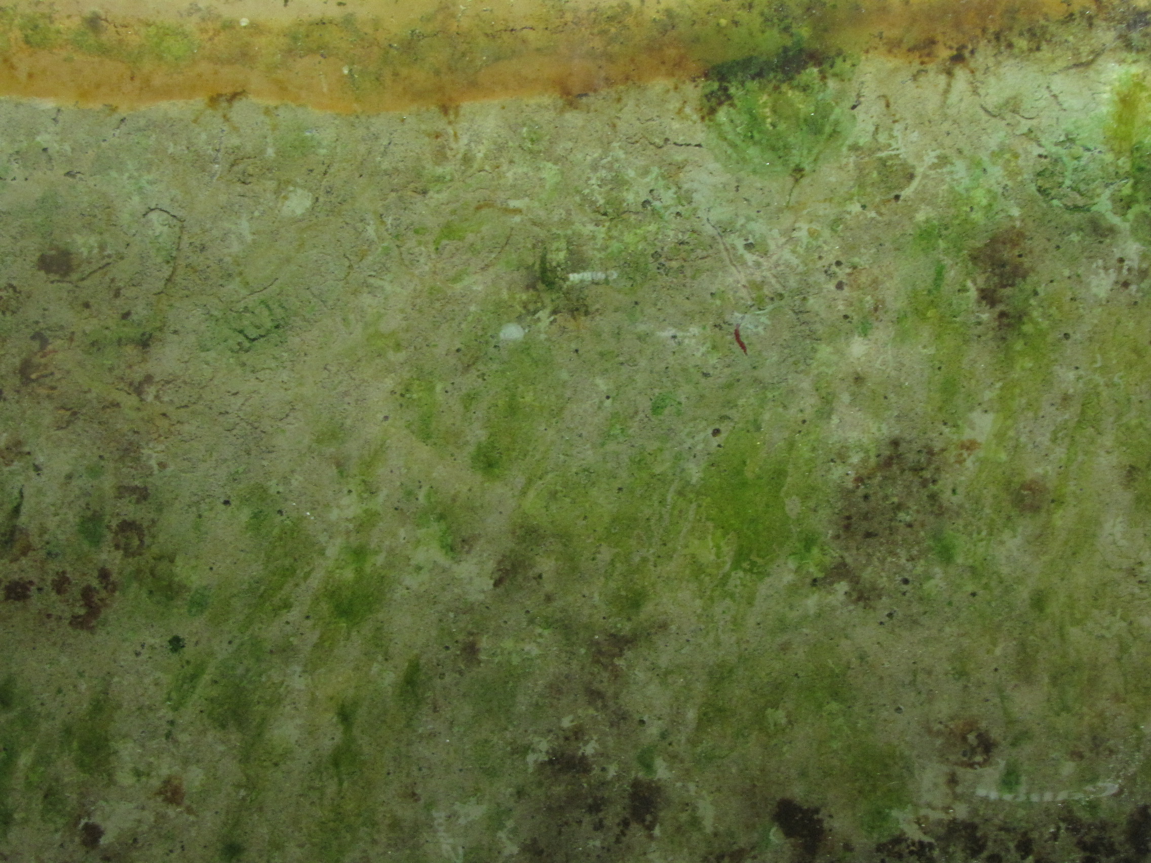
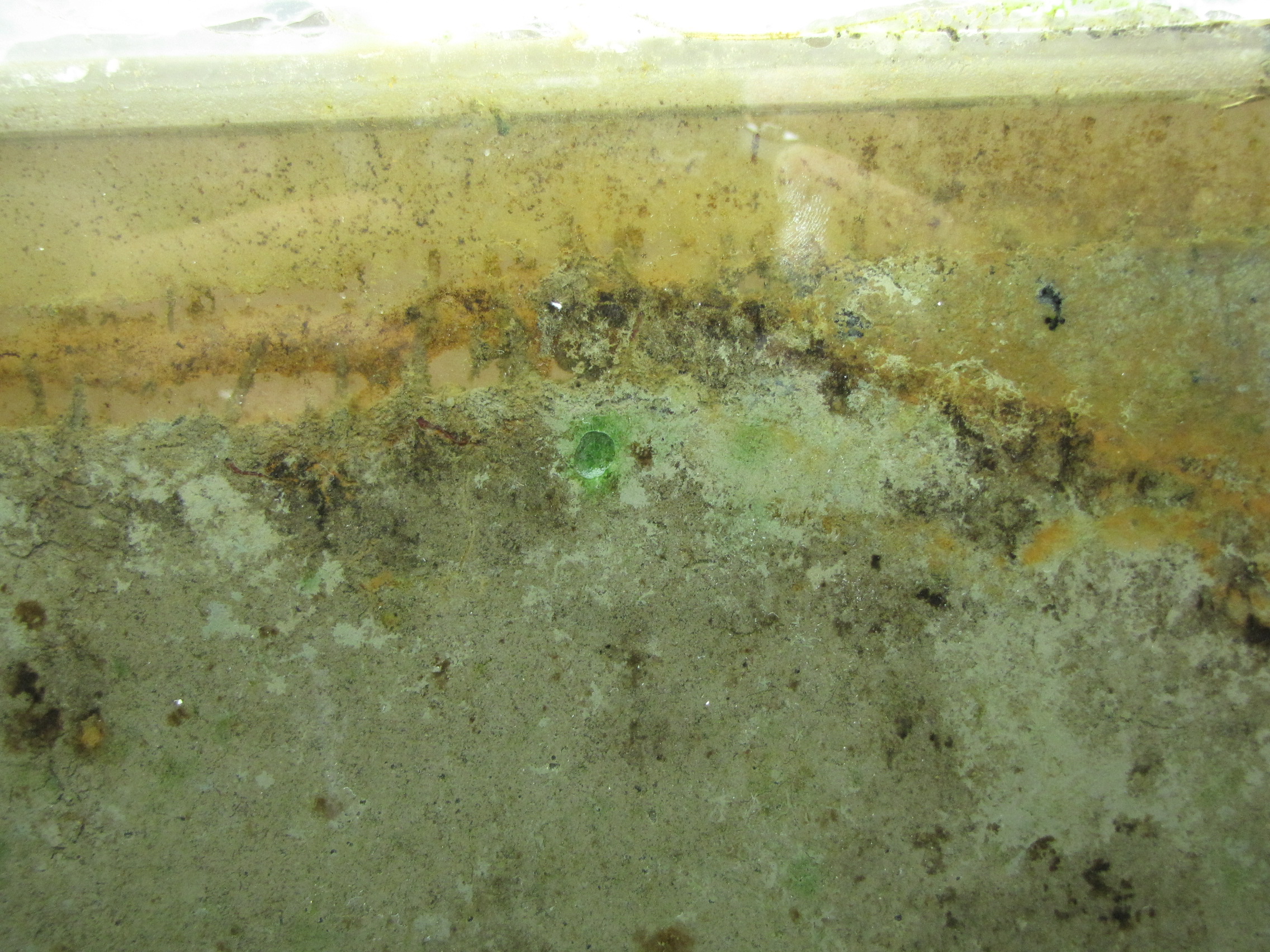
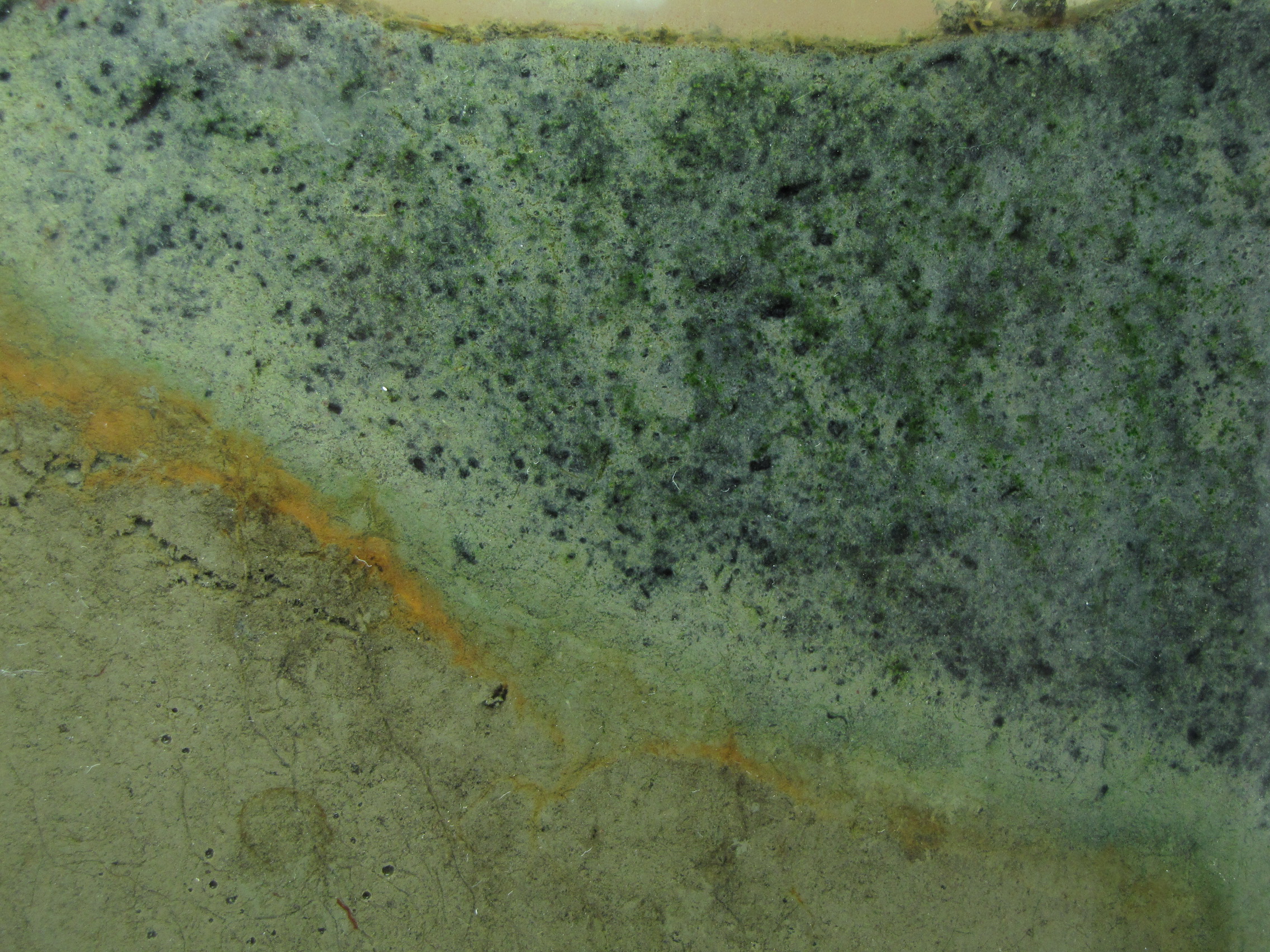
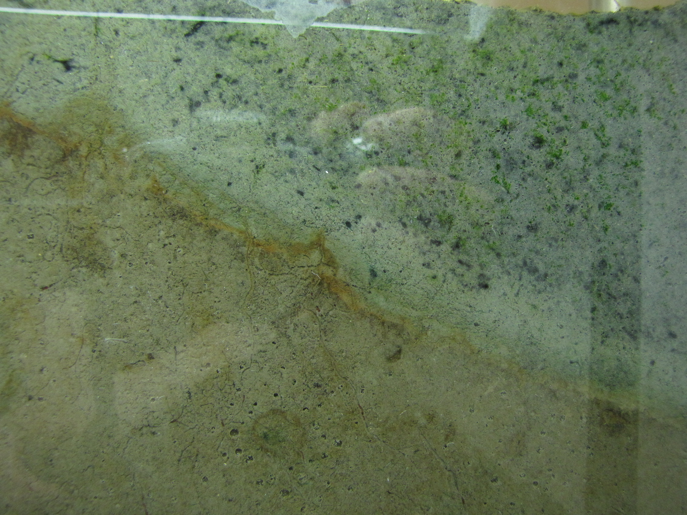
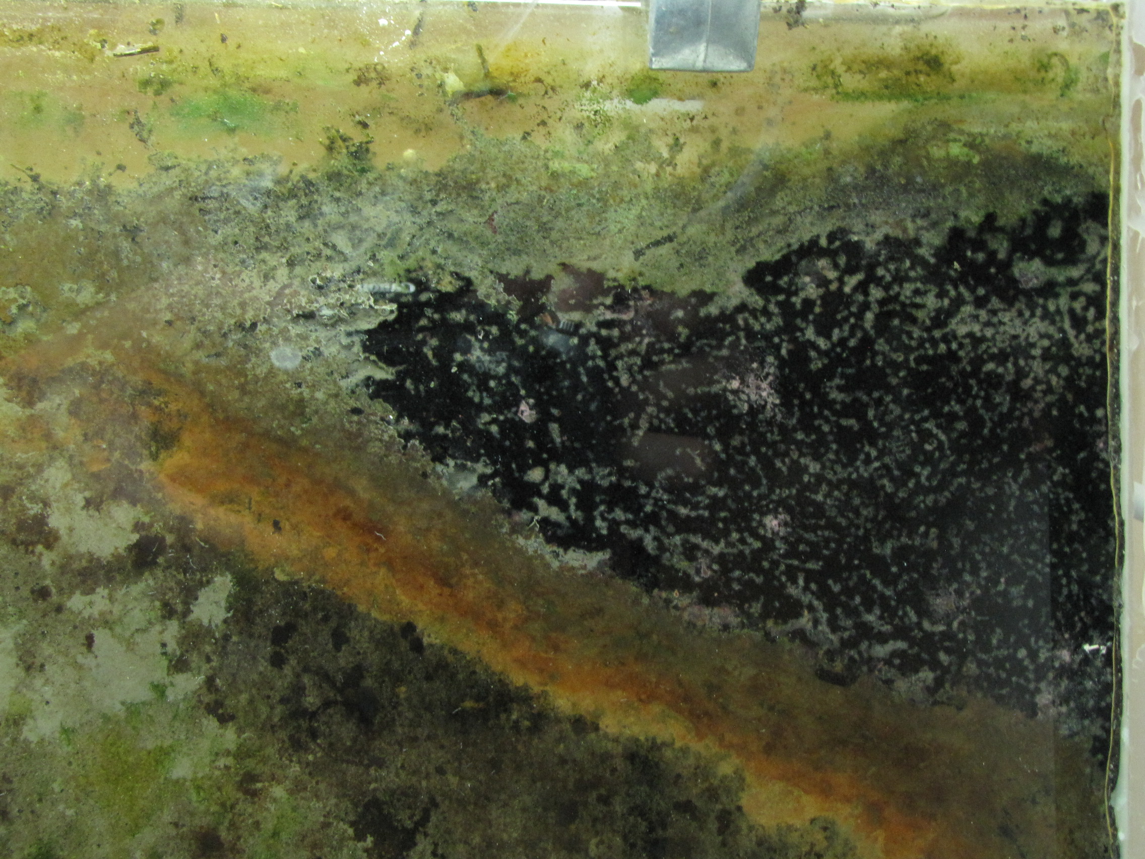
Close-up.  You can clearly see the layers of color starting to develop.  The whitish spots are actually gas bubbles, possibly CO2 or H2 produced from fermentation.  That CO2 will serve as a carbon source for autotrophs, and the H2 will serve as an energy source for hydrogenotrophs.  Some black specs are showing up in the modified mud.  Black is due to microbial production of hydrogen sulfide (H2S), which reacts with iron in the mud producing pyrite, which is black.

IMG_5903
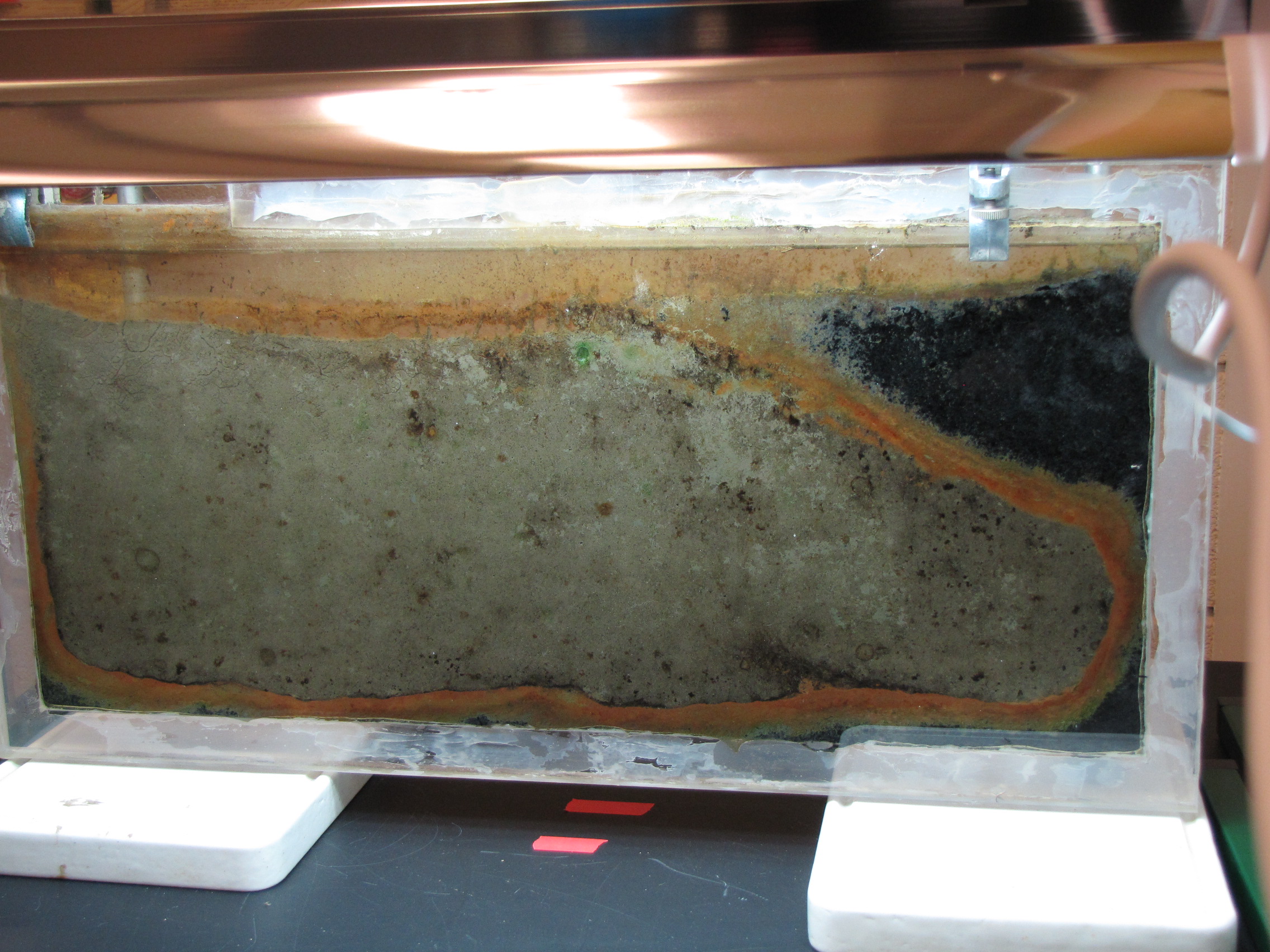
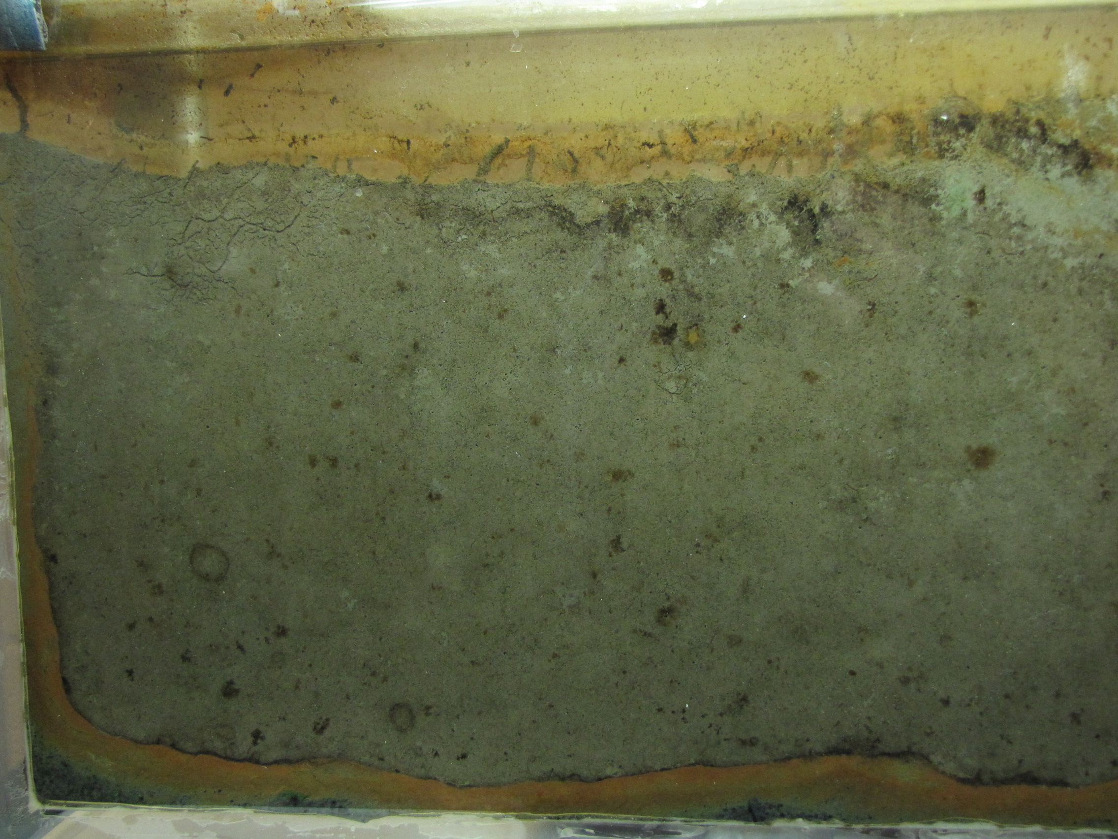
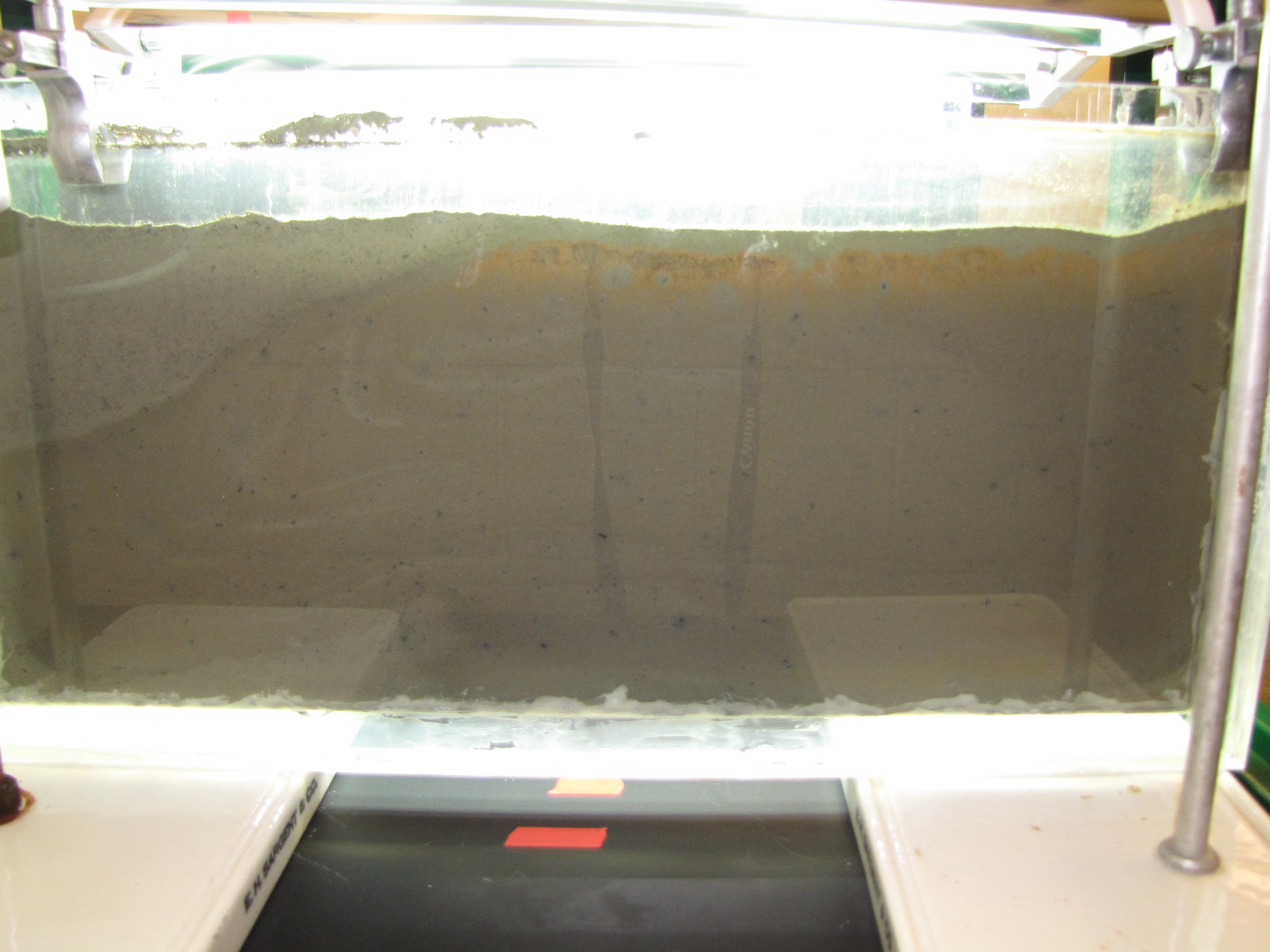
The back side of the panel. This side does not get direct light from the grow lamps. Some gets through on top and you can see the orange colored bacteria at the top. Note that the interface between the muds is less distinct here, showing that the colors on the illuminated side really are phototrophs.


Share