Tag Archives: green sulfur bacteria

Check out our newest paper in PLoS ONE!

Temporal and Spatial Distribution of the Microbial Community of Winogradsky Columns

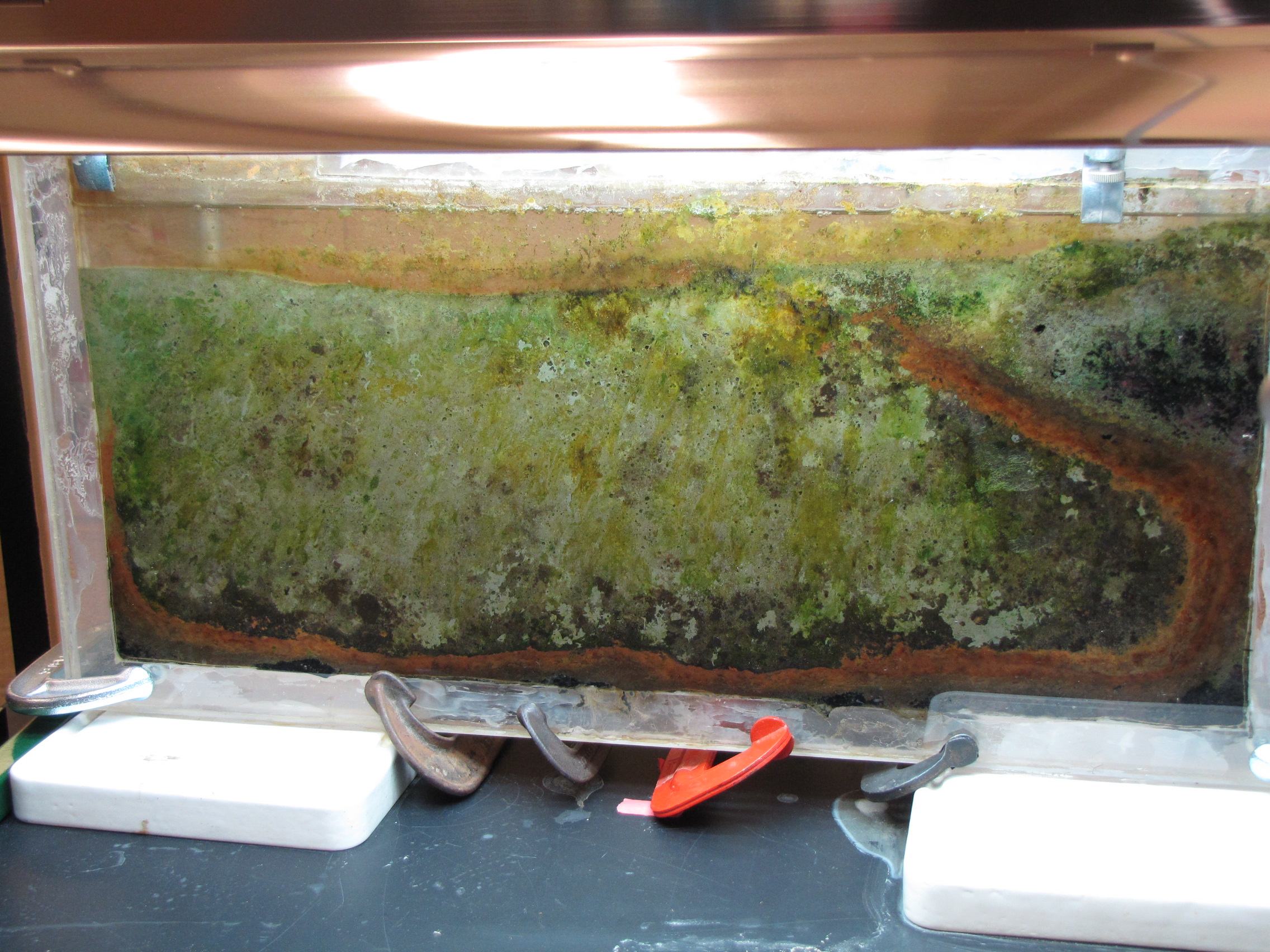
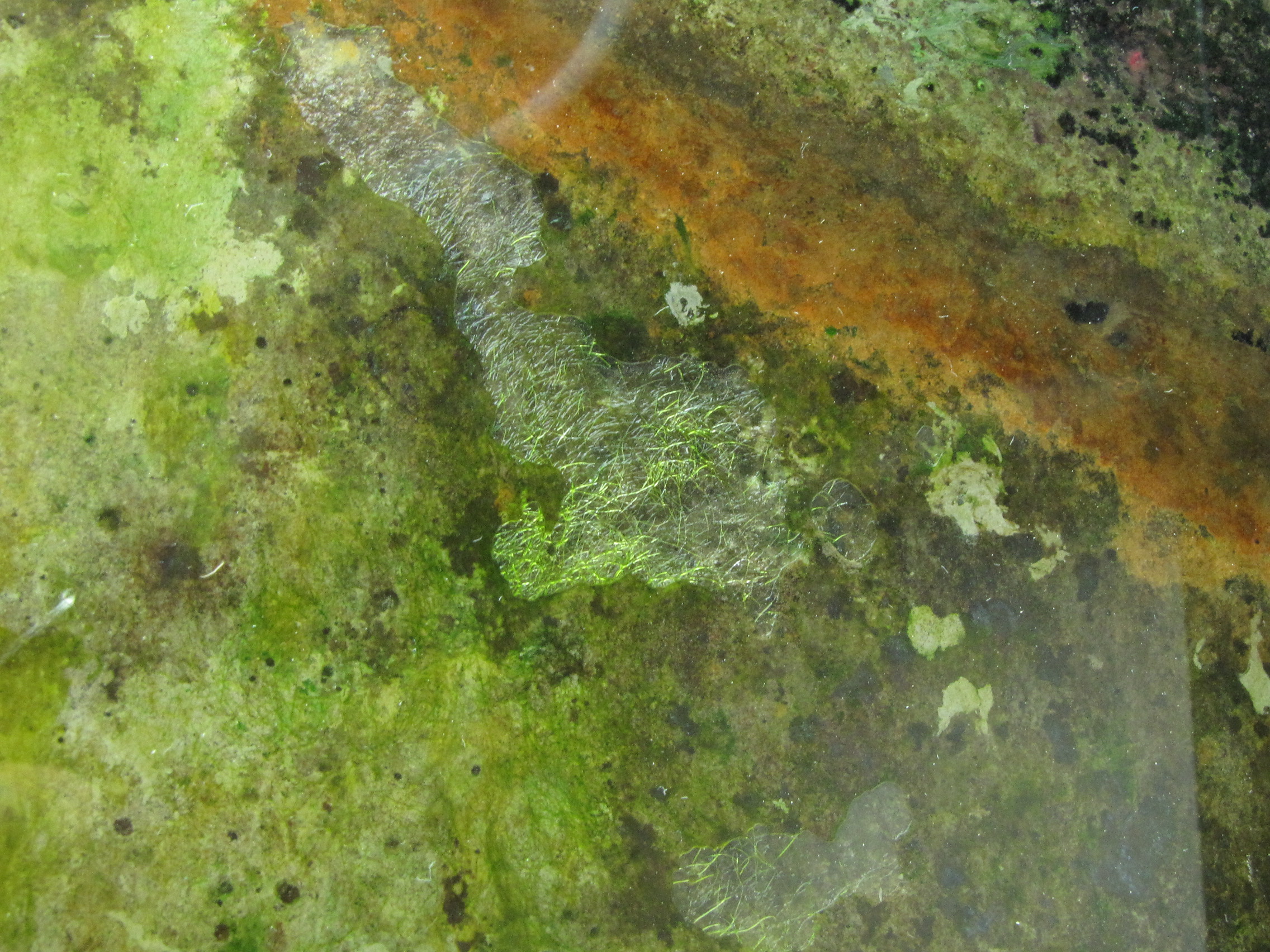
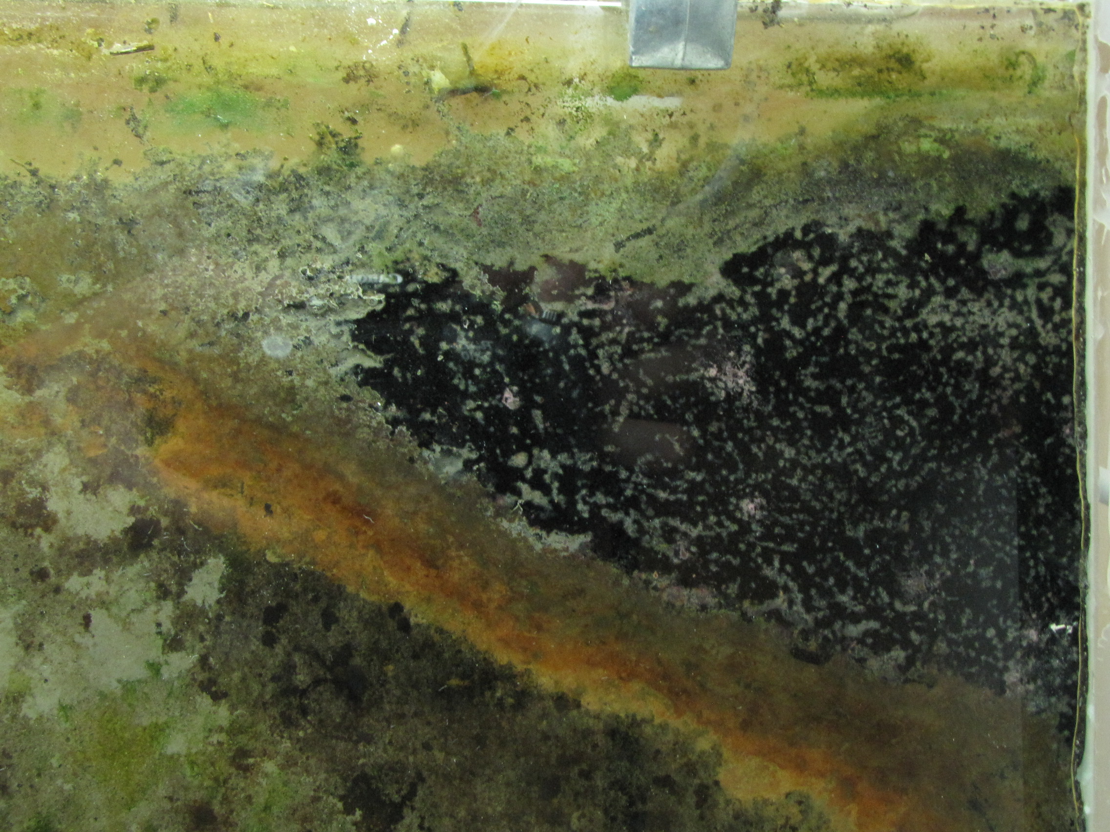
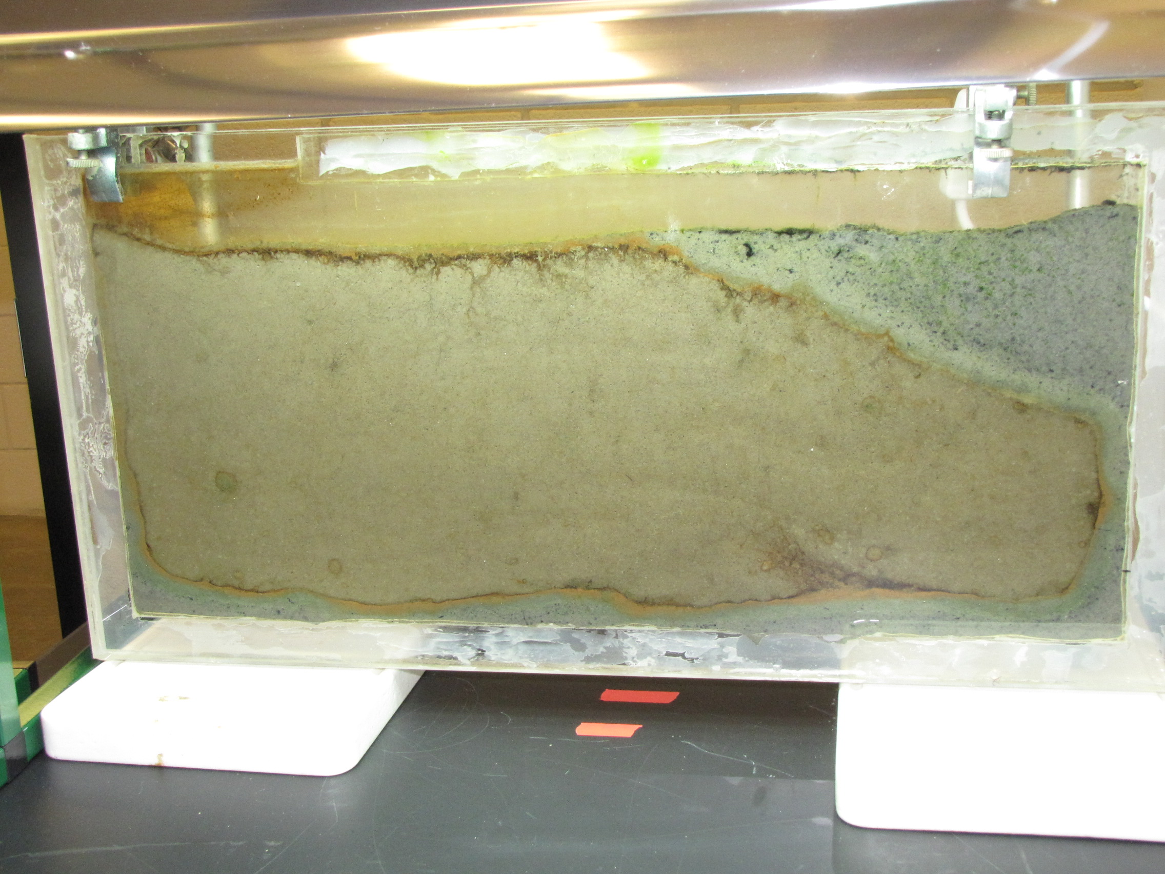
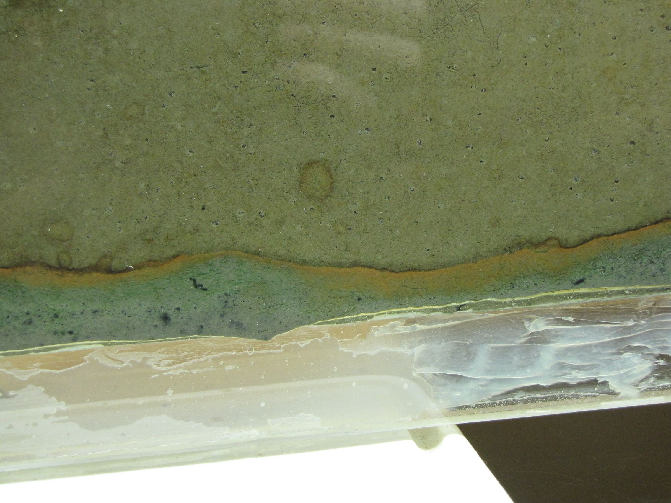
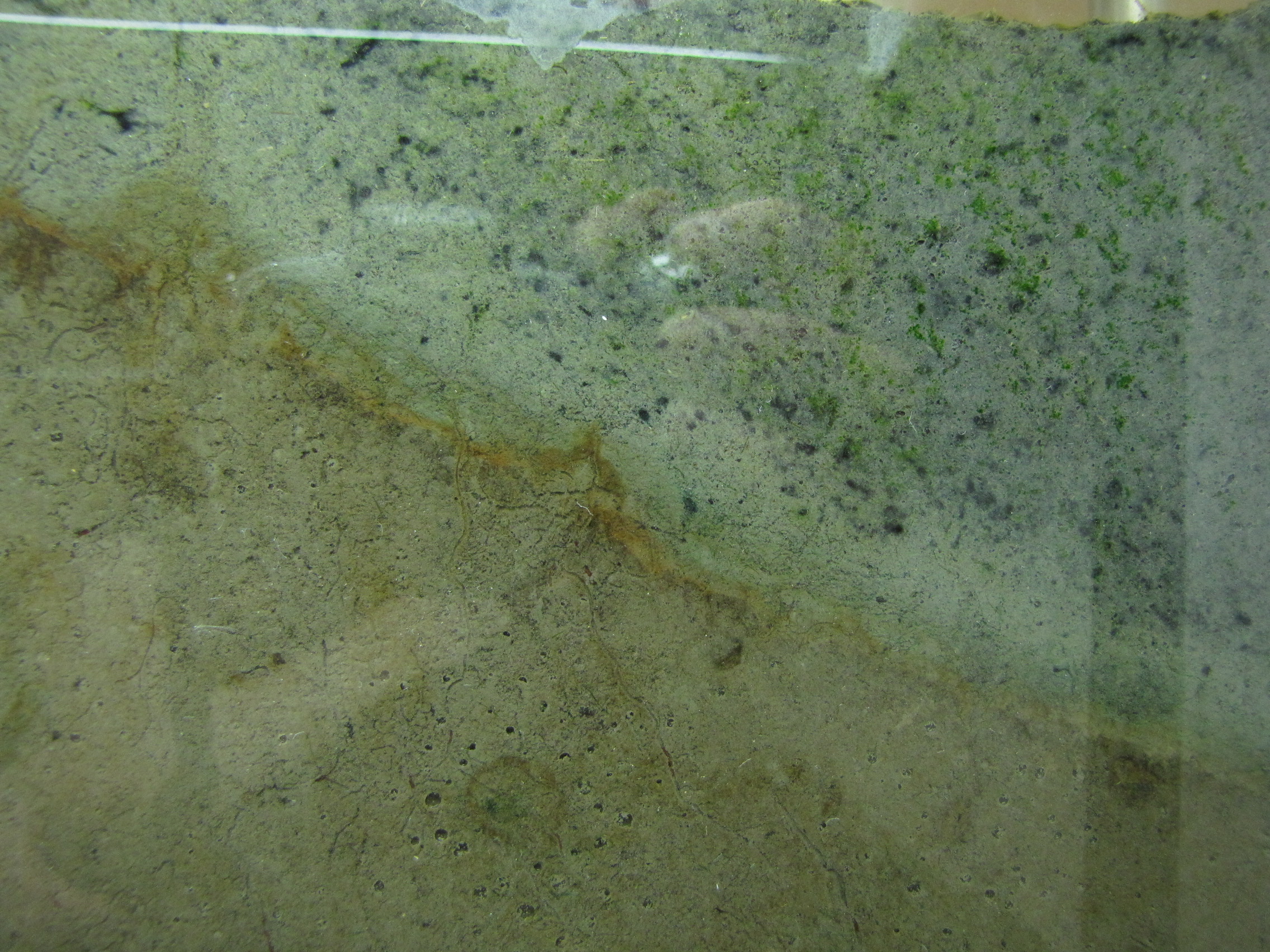
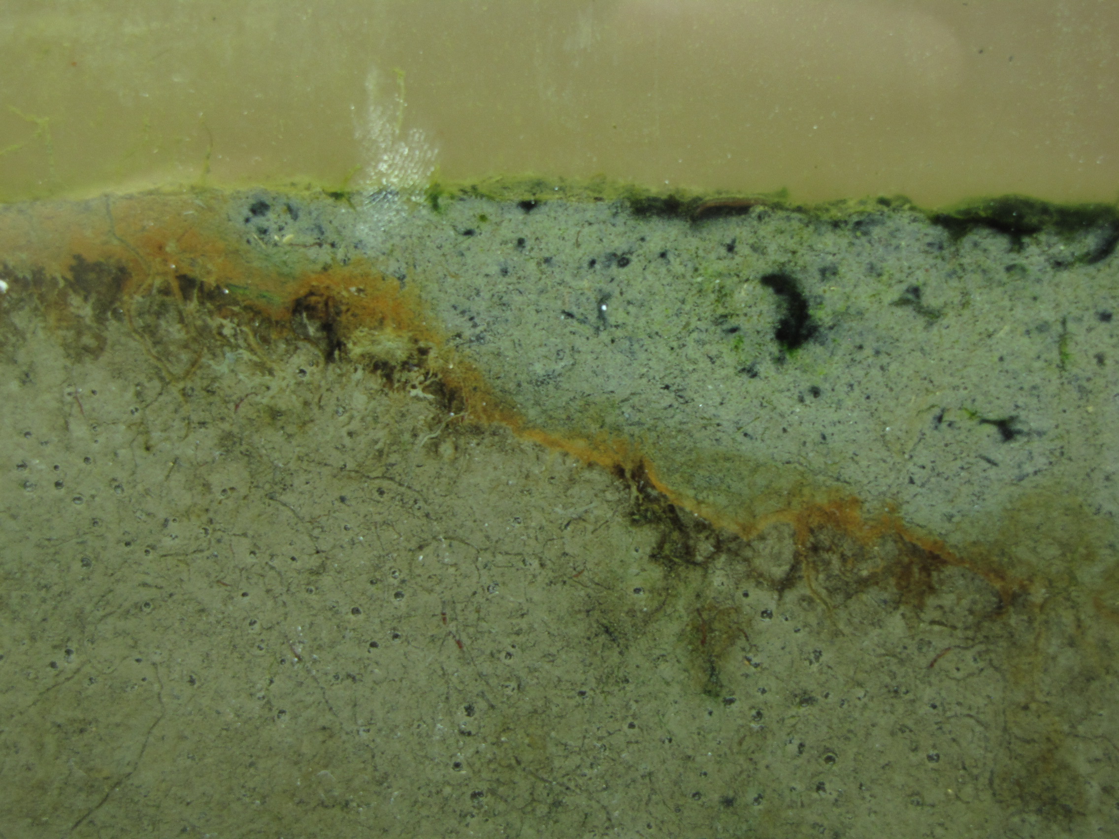
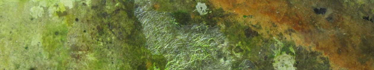
Winogradsky columns are model microbial ecosystems prepared by adding pond sediment to a clear cylinder with additional supplements and incubated with light. Environmental gradients develop within the column creating diverse niches that allow enrichment of specific bacteria. The enrichment culture can be used to study soil and sediment microbial community structure and function. In this study we used a 16S rRNA gene survey to characterize the microbial community dynamics during Winogradsky column development to determine the rate and extent of change from the source sediment community. Over a period of 60 days, the microbial community changed from the founding pond sediment population: Cyanobacteria, Chloroflexi, Nitrospirae, and Planctomycetes increased in relative abundance over time, while most Proteobacteria decreased in relative abundance. A unique, light-dependent surface biofilm community formed by 60 days that was less diverse and dominated by a few highly abundant bacteria. 67–72% of the surface community was comprised of highly enriched taxa that were rare in the source pond sediment, including the Cyanobacteria Anabaena, a member of the Gemmatimonadetes phylum, and a member of the Chloroflexi class Anaerolinea. This indicates that rare taxa can become abundant under appropriate environmental conditions and supports the hypothesis that rare taxa serve as a microbial seed bank. We also present preliminary findings that suggest that bacteriophages may be active in the Winogradsky community. The dynamics of certain taxa, most notably the Cyanobacteria, showed a bloom-and-decline pattern, consistent with bacteriophage predation as predicted in the kill-the-winner hypothesis. Time-lapse photography also supported the possibility of bacteriophage activity, revealing a pattern of colony clearance similar to formation of viral plaques. The Winogradsky column, a technique developed early in the history of microbial ecology to enrich soil microbes, may therefore be a useful model system to investigate both microbial and viral ecology.

Share

Watching Bacteria Grow: Winogradsky Panel Day 49

Various green bacteria are growing now all over the panel.  These are likely to be cyanobacteria and some green sulfur bacteria.

IMG_6151

You may also notice the addition of clamps along the bottom.  On Day 33, I arrived to find a puddle below the panel and all of the water drained out.  Luckily, the crack was too small to let any soil out.  The clamps are holding well so no more leaked out after I refilled it.  Perhaps I can claim that this is an experiment in shocking the ecosystem with a drying and aeration event?  I didn’t notice any significant changes in the following days and weeks, except the continued growth of green and yellow bacteria, so I suspect the shock wasn’t too detrimental.

IMG_6156

In the last several days, a filamentous green colony has developed.  Note the large gas bubbles around it.  This is likely a cyanobacteria like Oscillatoria, so the gas would be oxygen generated through its plant-like photosynthesis.  IMG_6120IMG_6141The two images above show the top right corner of the panel at day 35 and then day 49.  Notice how the black mud is slowly being covered by the growth of a very diverse mixture of microbes.

Share

Watching Bacteria Grow: Winogradsky Panel Day 4

The Winogradsky panel is developing much more quickly than I had expected. Differences are noticeable from day to day.

IMG_5928
The panel at day 4.

IMG_5933
The colors at the interface are richer and thicker. The green sulfur bacteria below the orange layer are more obvious now.

IMG_5937The modified mud layer now has more black spots and there are now some bright green spots, probably green sulfur bacteria.  GSB are more tolerant of high H2S concentrations than the purple sulfur bacteria, so are growing well in the areas where the black spots tell us there is H2S.  Note the gas bubbles at the bottom center of the image.  The squiggly lines are actually paths left behind by the movement of small, red larvae of midge flies, called blood worms.  They made their way up to the top, and are now all apparently dead.  (And will be food for some bacteria).

IMG_5944


Share