
Check out the paper in PLoS ONE!
Ethan Rundell ’13 worked in my lab for two years, his work culminating in first authorship on this paper.
Abstract
A Winogradsky column is a clear glass or plastic column filled with enriched sediment. Over time, microbial communities in the sediment grow in a stratified ecosystem with an oxic top 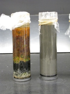 layer and anoxic sub-surface layers. Winogradsky columns have been used extensively to demonstrate microbial nutrient cycling and metabolic diversity in undergraduate microbiology labs. In this study, we used high-throughput 16s rRNA gene sequencing to investigate the microbial diversity of Winogradsky columns. Specifically, we tested the impact of sediment source, supplemental cellulose source, and depth within the column, on microbial community structure. We found that the Winogradsky columns were highly diverse communities but are dominated by three phyla: Proteobacteria, Bacteroidetes, and Firmicutes. The community is structured by a founding population dependent on the source of sediment used to prepare the columns and is differentiated by depth within the column. Numerous biomarkers were identified distinguishing sample depth, including Cyanobacteria, Alphaproteobacteria, and Betaproteobacteria as biomarkers of the soil-water interface, and Clostridia as a biomarker of the deepest depth. Supplemental cellulose source impacted community structure but less strongly than depth and sediment source. In columns dominated by Firmicutes, the family Peptococcaceae was the most abundant sulfate reducer, while in columns abundant in Proteobacteria, several Deltaproteobacteria families, including Desulfobacteraceae, were found, showing that different taxonomic groups carry out sulfur cycling in different columns. This study brings this historical method for enrichment culture of chemolithotrophs and other soil bacteria into the modern era of microbiology and demonstrates the potential of the Winogradsky column as a model system for investigating the effect of environmental variables on soil microbial communities.
Rundell EA, Banta LM, Ward DV, Watts CD, Birren B, Esteban, DJ. (2014) 16S rRNA Gene Survey of Microbial Communities in Winogradsky Columns. PLoS ONE 9(8): e104134. doi:10.1371/journal.pone.0104134
