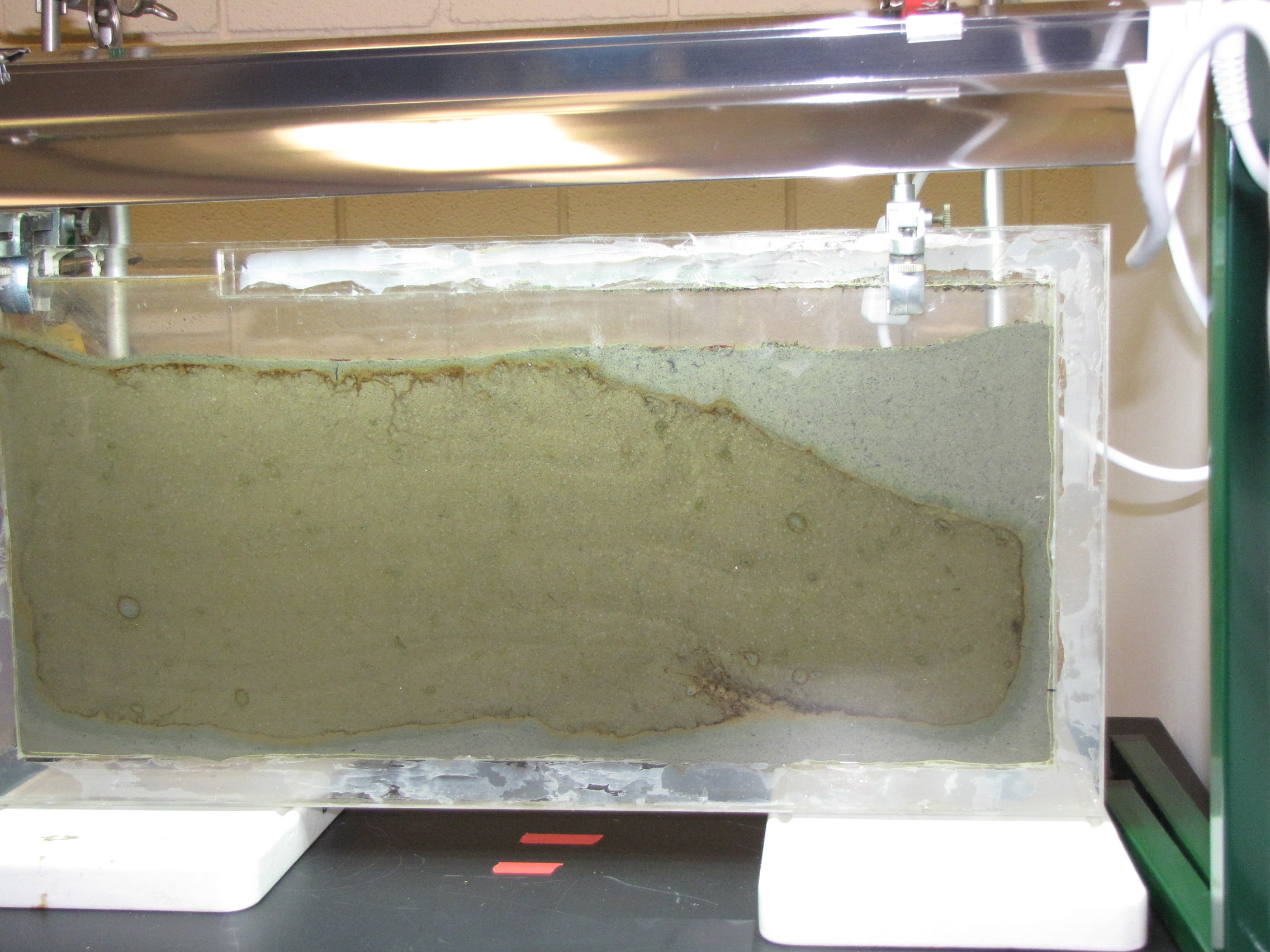
Watching Bacteria Grow: Winogradsky Panel Day 7

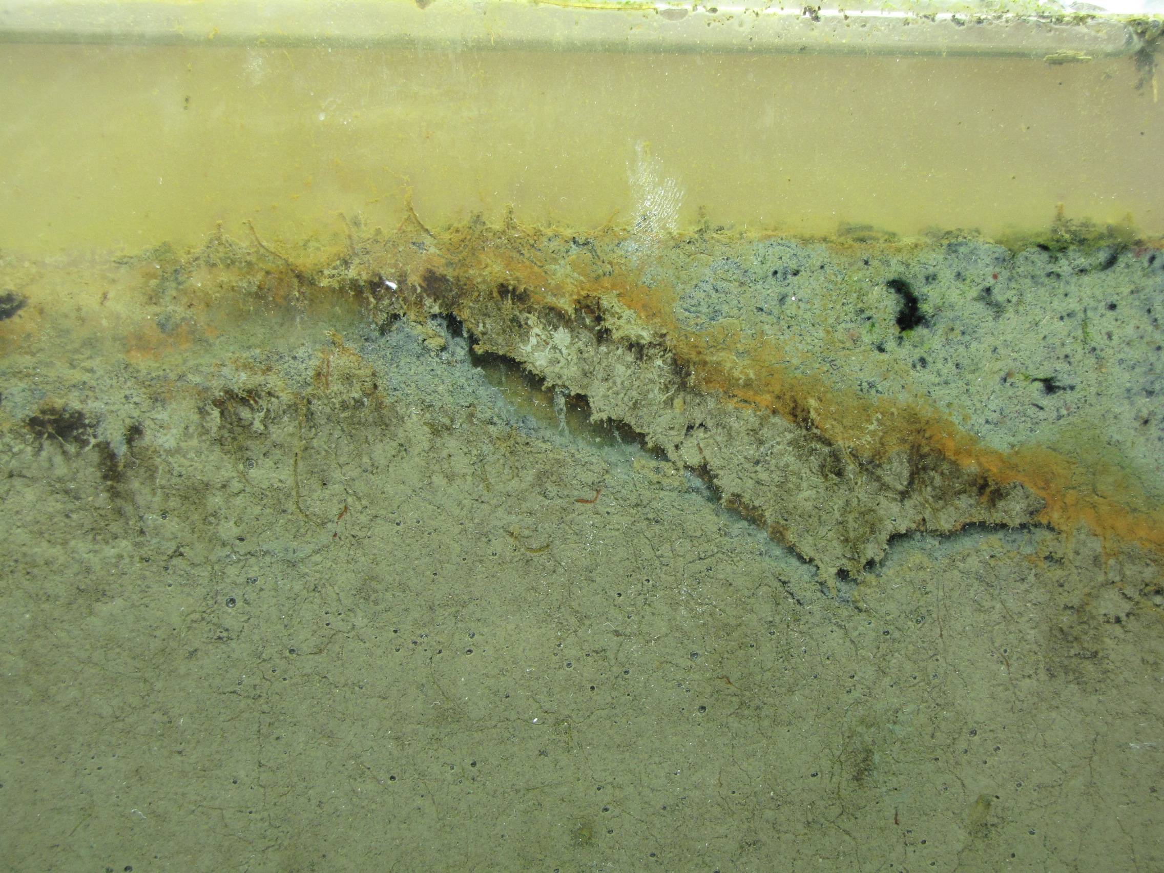
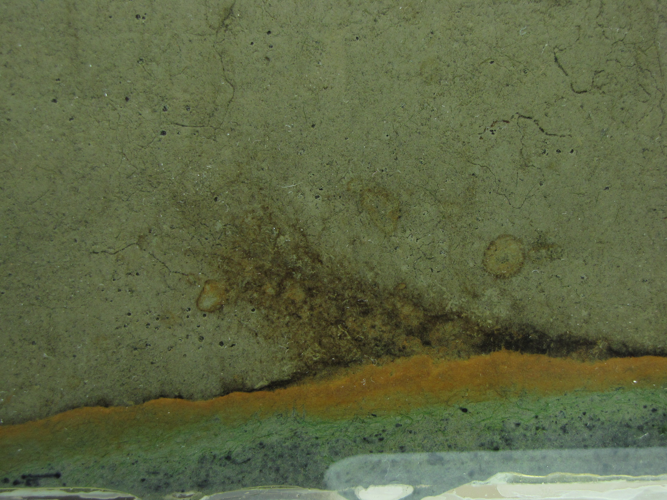
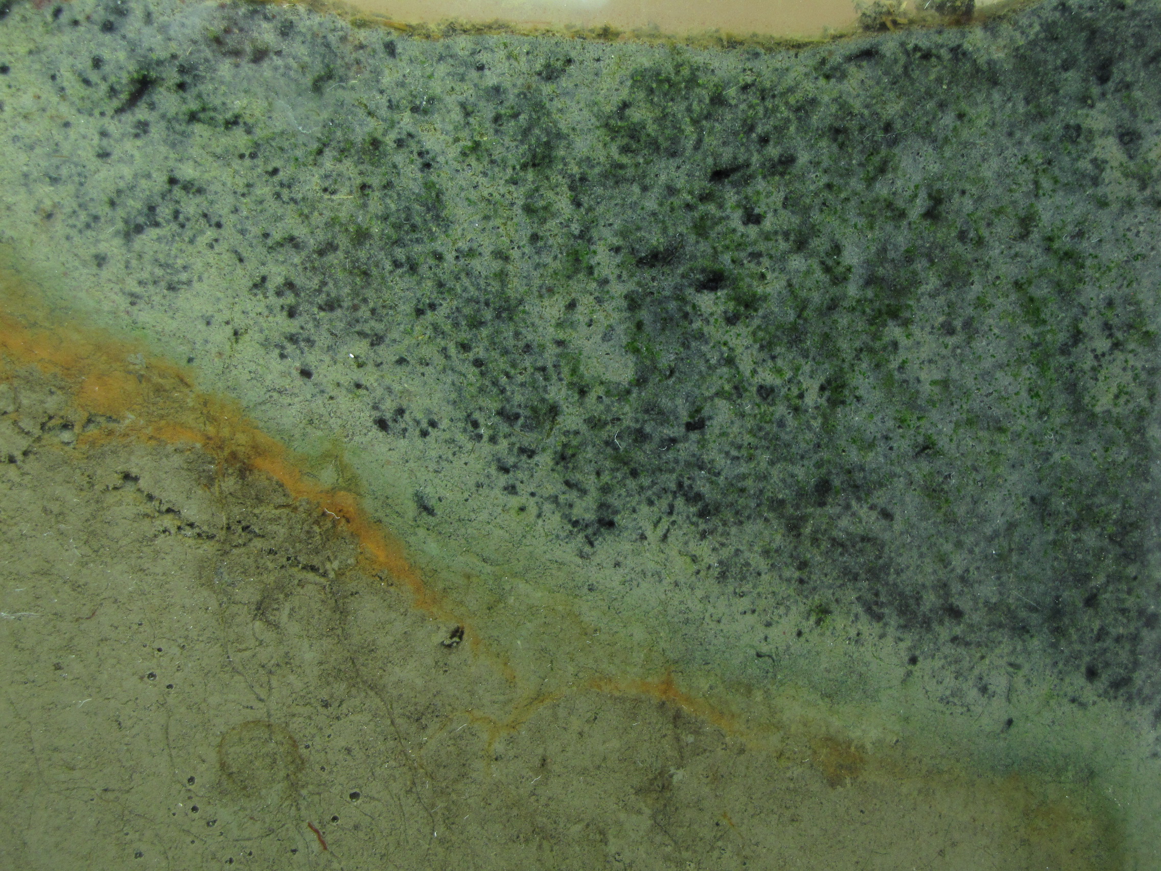
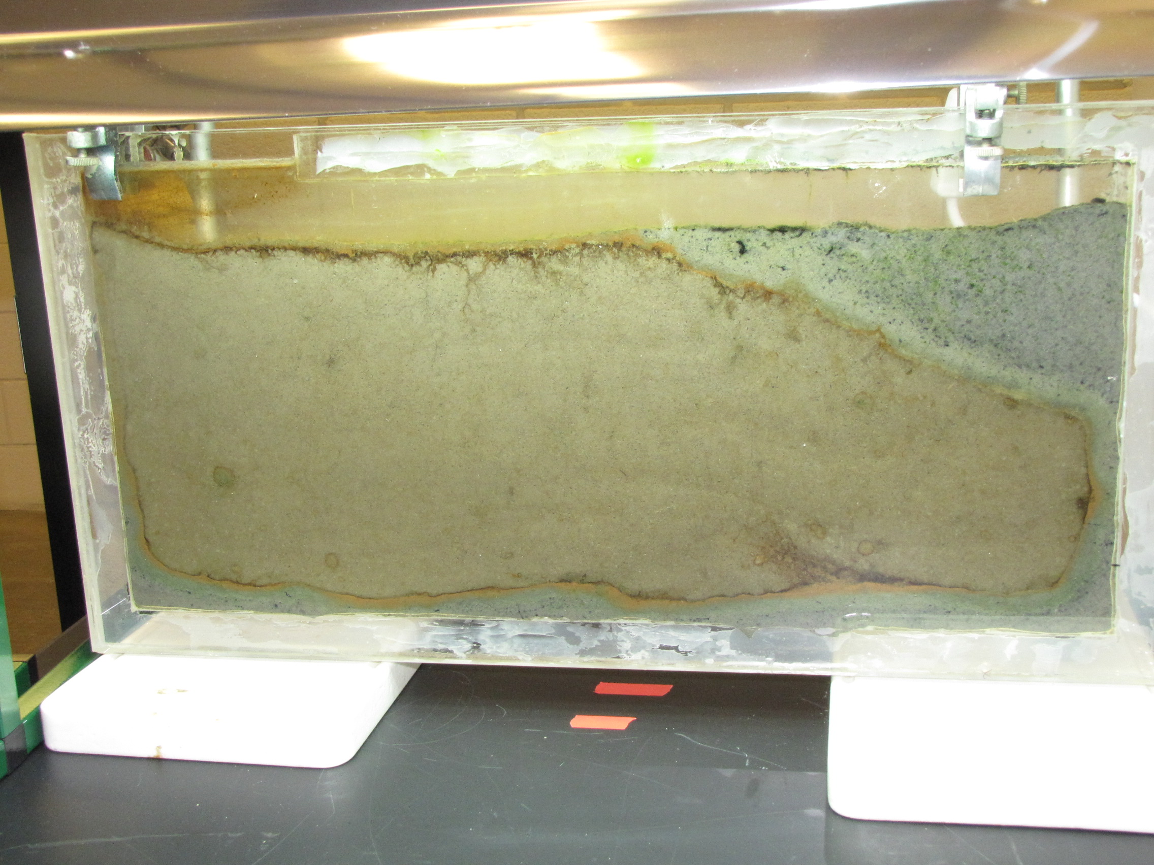
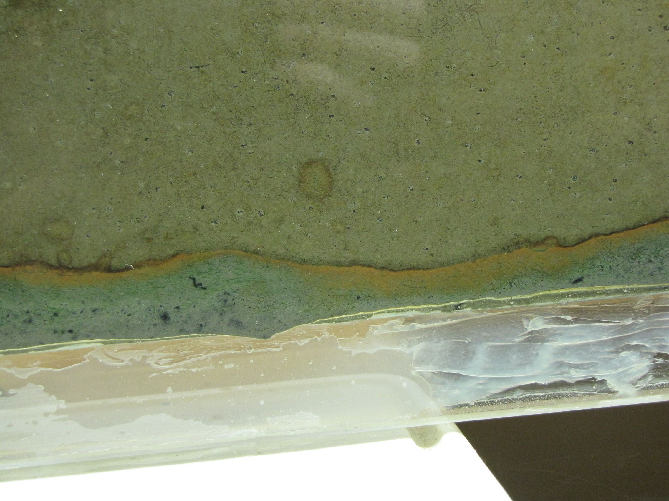
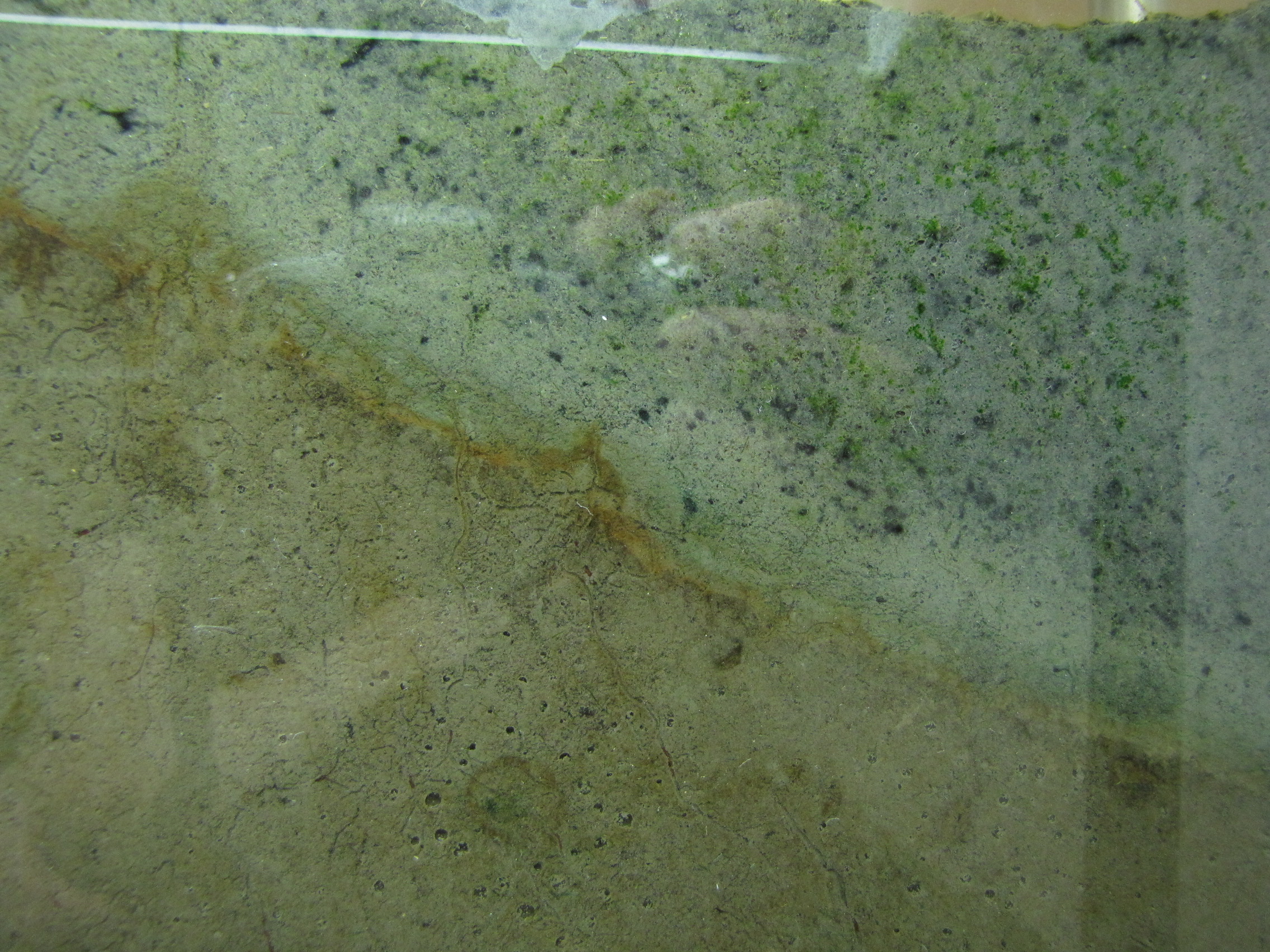
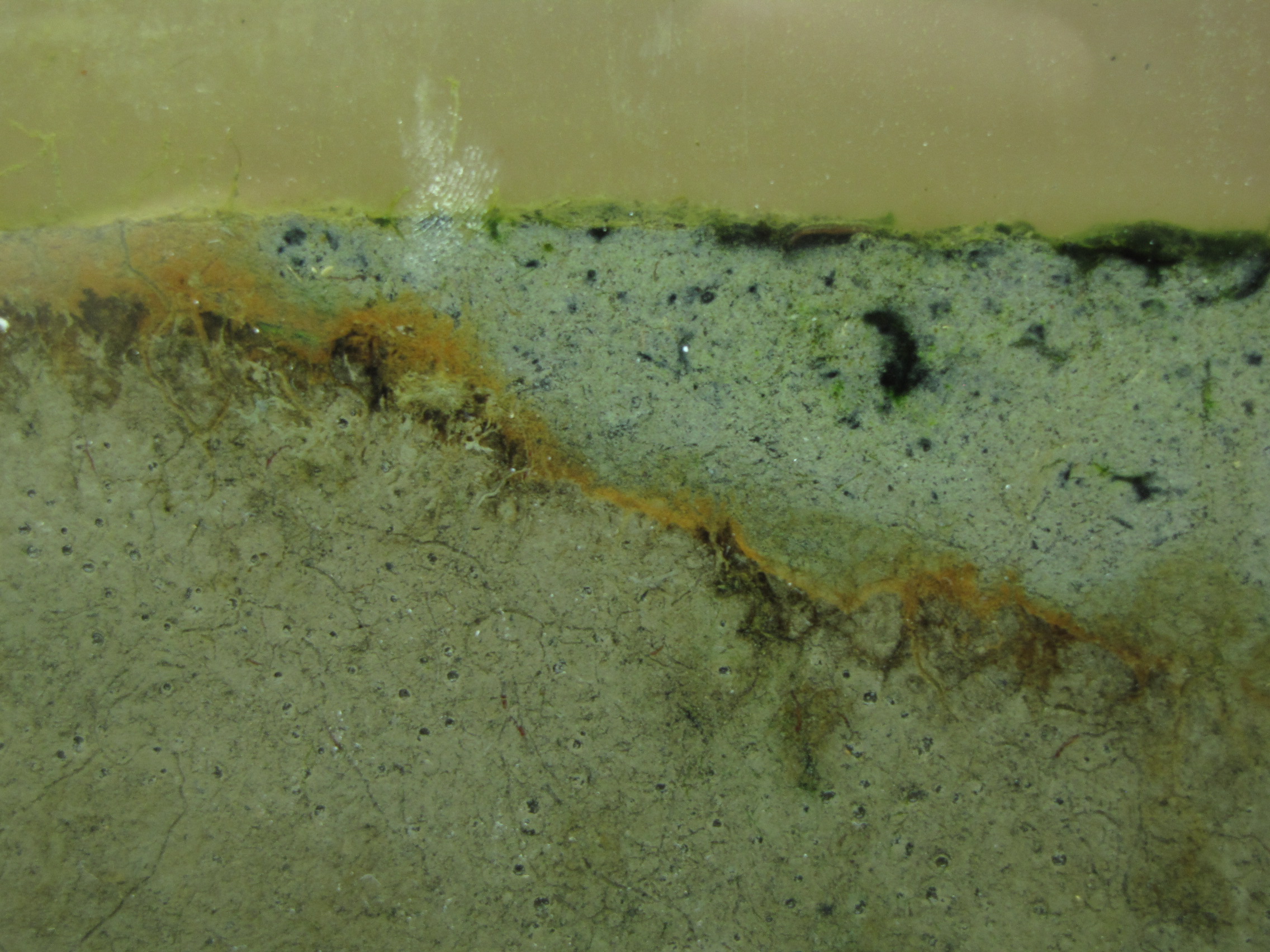
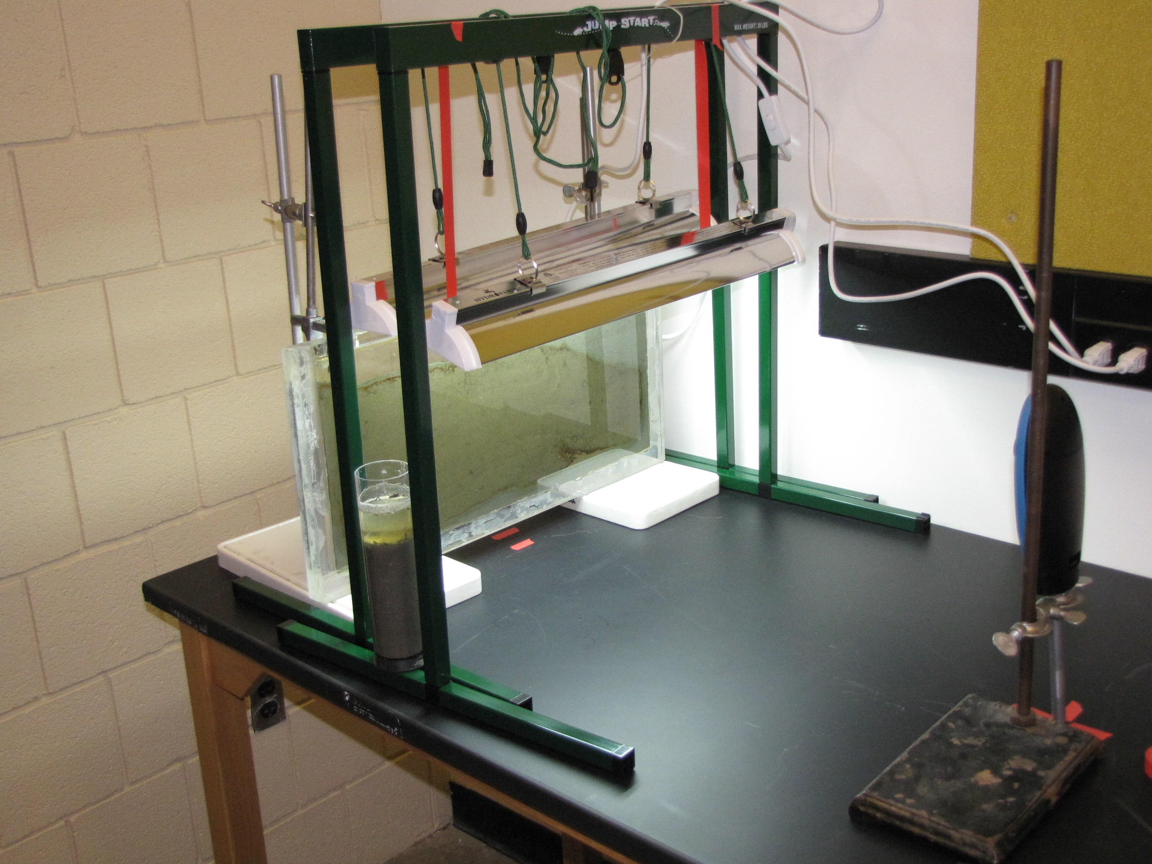
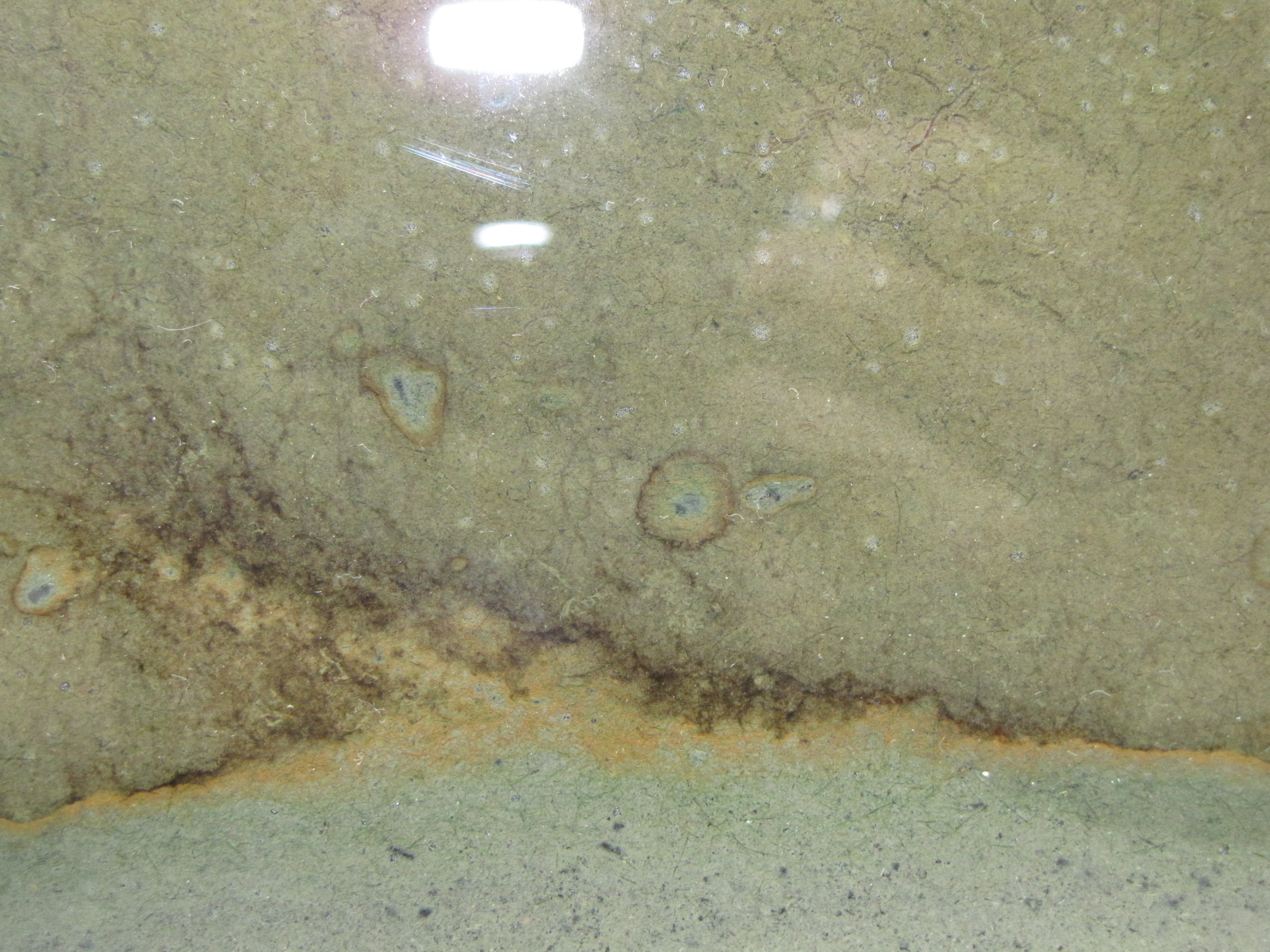
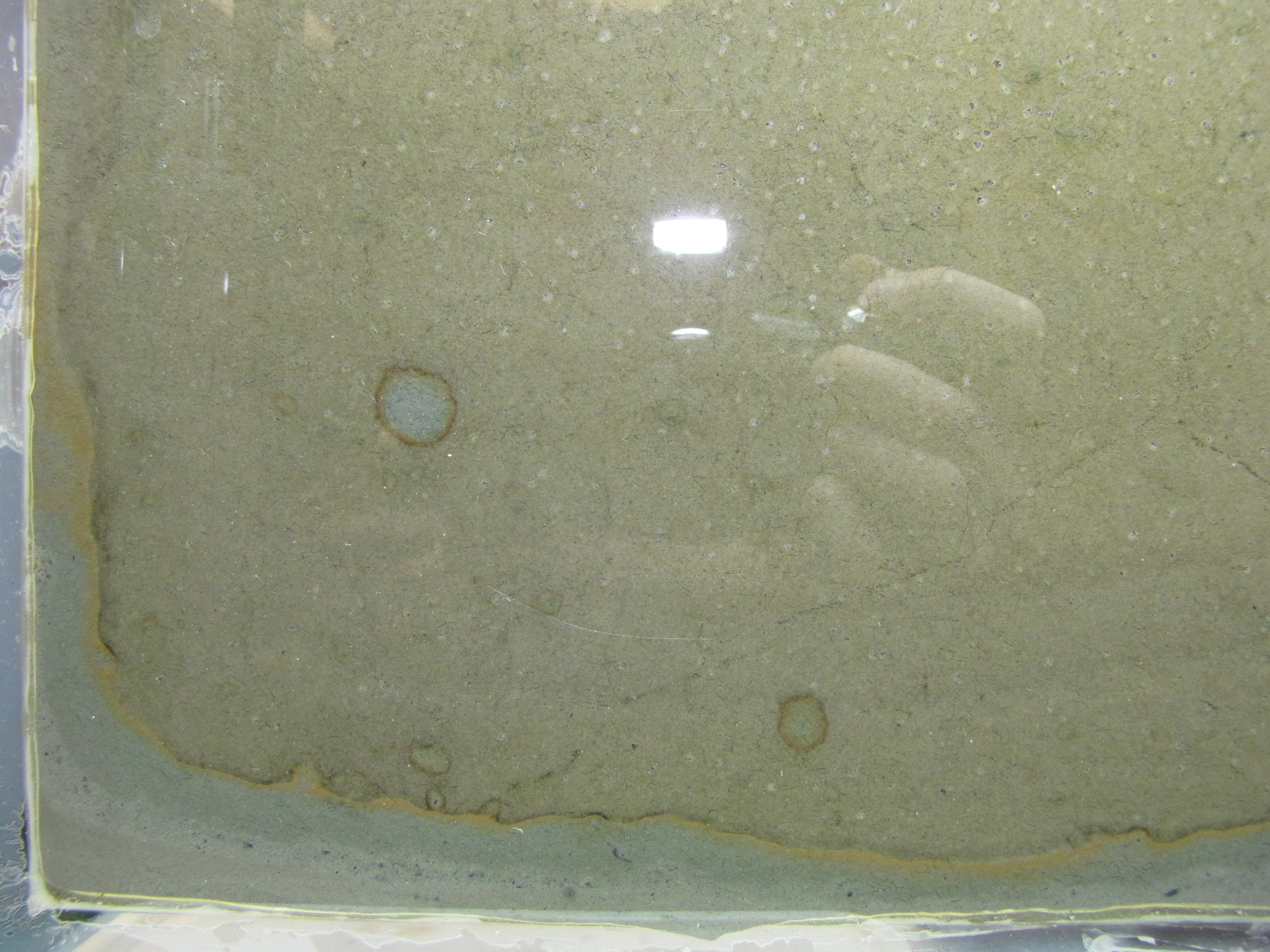
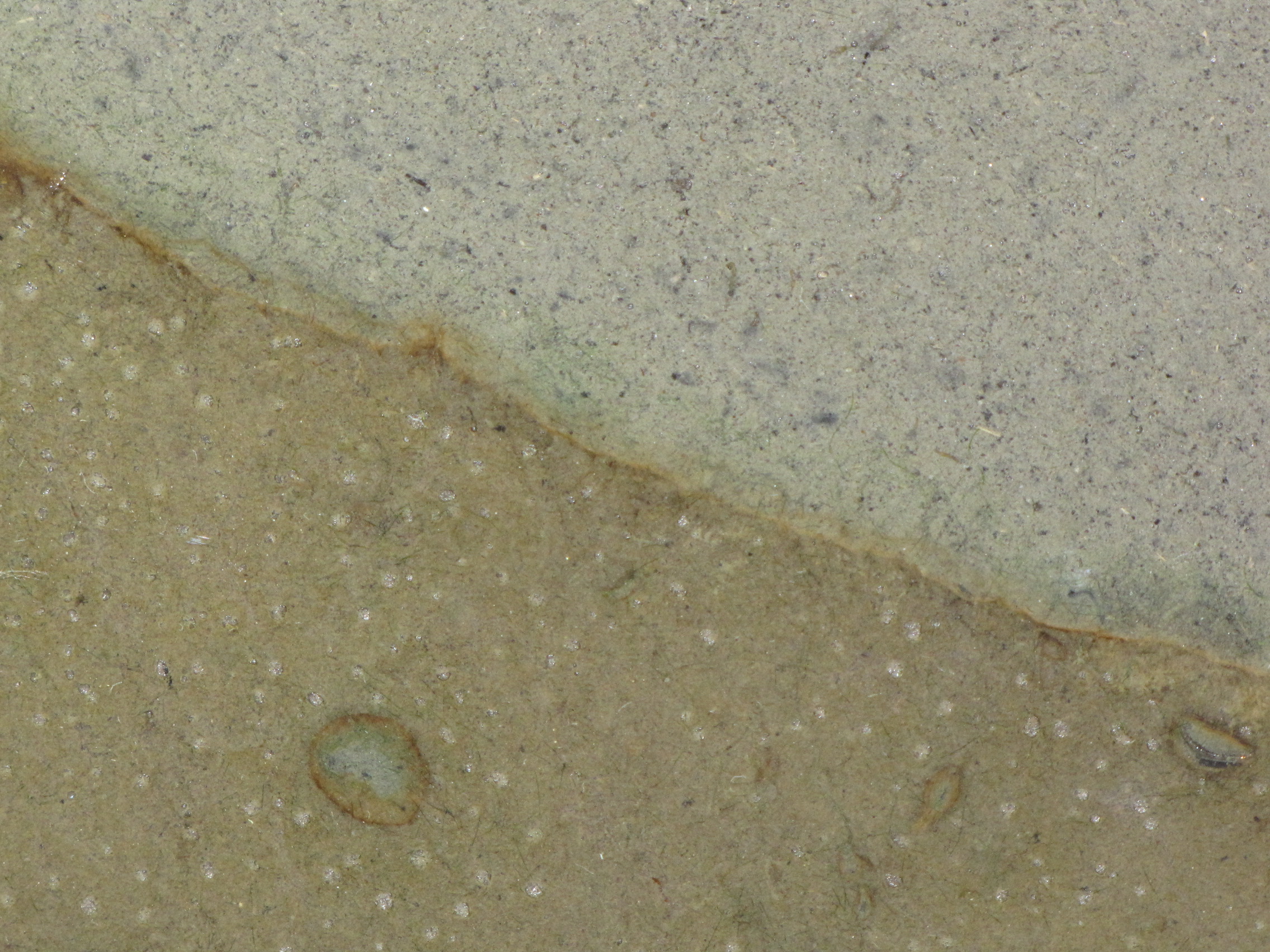
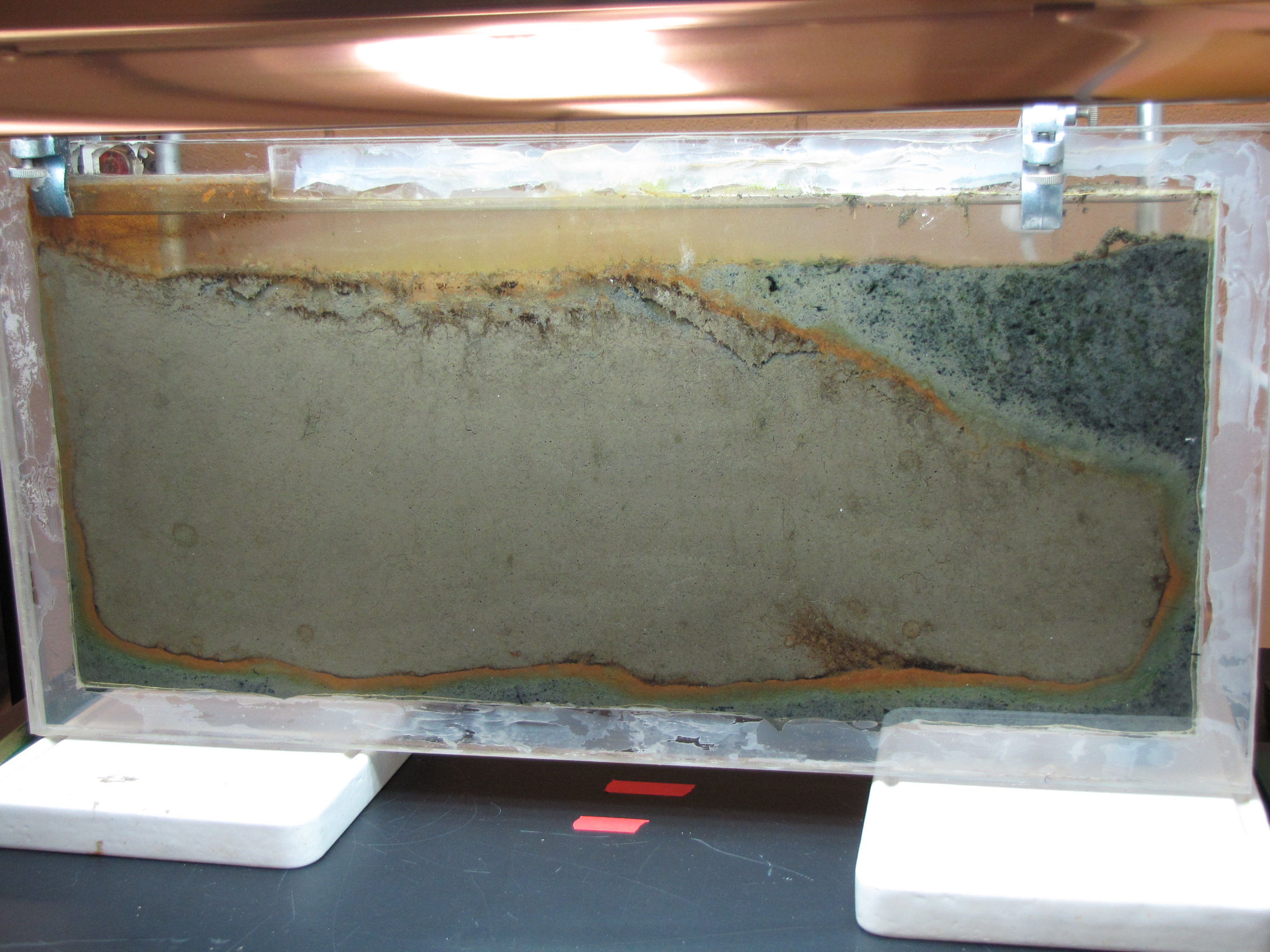
The panel is changing more slowly now, but the oranges and greens are getting richer and the sulfur-enriched mud is showing more and coalescing black spots.
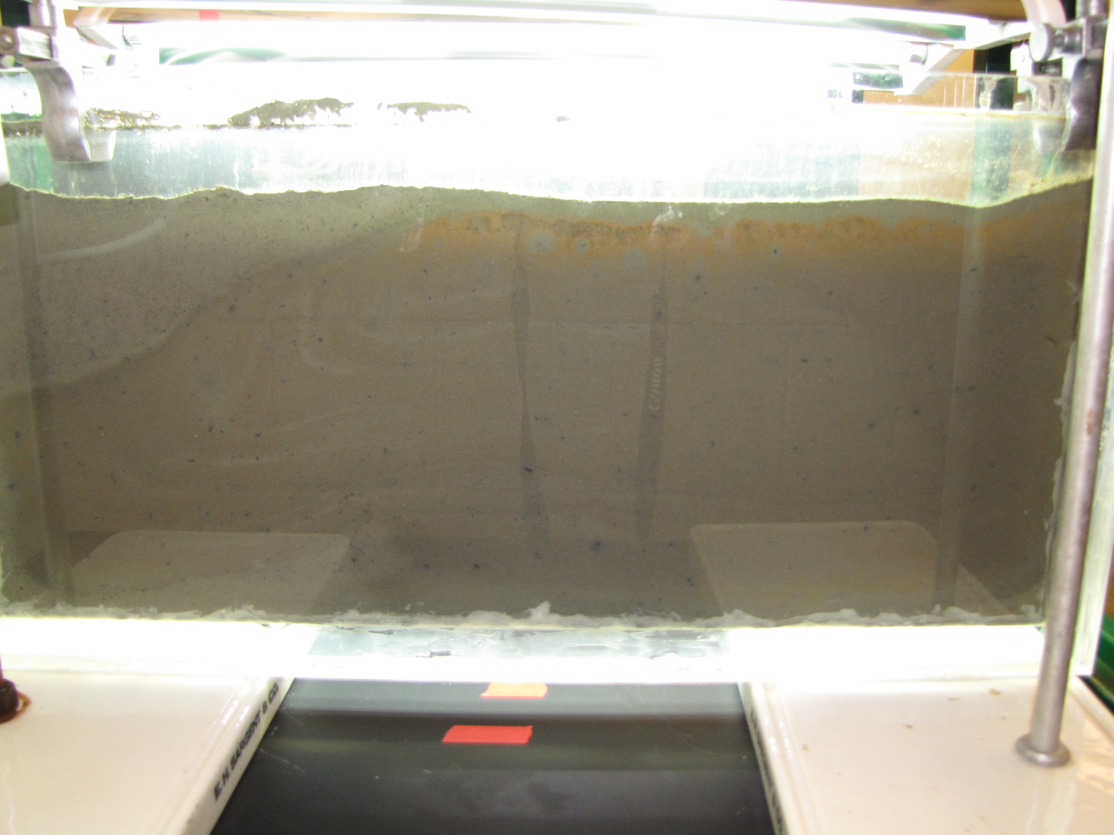
IMG_5962The level of the mud dropped overnight.  This could be due to settling of the mud and/or release of gas bubbles.  There are some water-filled gaps, and the water level is lower.  There is now air above the entire length of the water surface, instead of just at the opening of the panel (top left).  Note that there is also a left to right gradient in the water, which was already present before settling.  Near the opening, there is brownish/red colonies growing on the walls, probably aerobes (requires oxygen) explaining why its not present further from the opening.  The water is murky from left to right, perhaps microbes that can tolerate low levels of oxygen, up to a point near the right side where the water layer is still clear.

IMG_5965
IMG_5967
IMG_5968


Share

Watching Bacteria Grow: Winogradsky Panel Day 4

The Winogradsky panel is developing much more quickly than I had expected. Differences are noticeable from day to day.

IMG_5928
The panel at day 4.

IMG_5933
The colors at the interface are richer and thicker. The green sulfur bacteria below the orange layer are more obvious now.

IMG_5937The modified mud layer now has more black spots and there are now some bright green spots, probably green sulfur bacteria.  GSB are more tolerant of high H2S concentrations than the purple sulfur bacteria, so are growing well in the areas where the black spots tell us there is H2S.  Note the gas bubbles at the bottom center of the image.  The squiggly lines are actually paths left behind by the movement of small, red larvae of midge flies, called blood worms.  They made their way up to the top, and are now all apparently dead.  (And will be food for some bacteria).

IMG_5944


Share

Watching Bacteria Grow: Winogradsky Panel

One of the research projects in my lab is to investigate the microbial communities present in Winogradsky columns. You can read more about what a Winogradsky column is here.  Over the next little while, I will be carefully watching the development of the microbial communities over time.  This will help me set up a future experiment in which I will take samples over time and determine what bacteria are present and at what abundance to see how the community changes.  I will be posting regular updates as the Winogradsky panel develops.

IMG_5882
The set-up: the panel is 1′ x 2′ plexiglass with a 1/4″ gap filled with mud from a pond on Vassar campus, grow lamps, and time-lapse camera.

IMG_5883
The panel after 2 days. The panel was prepared by adding calcium carbonate, calcium sulfate, and dried leaves to mud from a pond on Vassar campus.  We sifted the mud to make a fine slurry.  We collected leaves from near the pond and baked them to dry them, then blended in a blender to make as fine a powder as possible.  The leaves serve as a source of cellulose.  This supplemented mud was poured to a depth of about 4 cm.  Then we poured unmodified mud (no additives) on top.  Instead of just layering on top though, the bottom layer was displaced, and most of it was pushed to the right side and up to the top.  You can see it clearly here: the mud with additives has the lighter grey color.

Note the orange and brownish-orange along the junction between the two muds. This is microbial growth, probably the phototrophic “purple bacteria.” Purple bacteria come in many colors, including red, orange, brown, and yes, purple.  They use light for energy but unlike plants they use H2S instead of water (H2O) as an electron source, and produce sulfur instead of oxygen.  You can also see it in the little circles where a spot of sulfur-enriched mud got surrounded by unmodified mud.

IMG_5886Close-up of the layers. There is a faint hint of green below the orange layer, these are green sulfur bacteria.

IMG_5898
Another close-up.

IMG_5884Close-up.  You can clearly see the layers of color starting to develop.  The whitish spots are actually gas bubbles, possibly CO2 or H2 produced from fermentation.  That CO2 will serve as a carbon source for autotrophs, and the H2 will serve as an energy source for hydrogenotrophs.  Some black specs are showing up in the modified mud.  Black is due to microbial production of hydrogen sulfide (H2S), which reacts with iron in the mud producing pyrite, which is black.

IMG_5903The back side of the panel. This side does not get direct light from the grow lamps. Some gets through on top and you can see the orange colored bacteria at the top. Note that the interface between the muds is less distinct here, showing that the colors on the illuminated side really are phototrophs.


Share

Your personal genome sequence

As a bonus question on their final exam, I asked my intro biology students whether or not they would want to have their genome sequenced. The question was:

It is already possible to get your whole genome sequenced and it wont be long before it becomes a routine part of your medical care. Having your genome sequenced can potentially reveal information like increased or decreased susceptibility to specific viral diseases, relative likelihood of developing diseases like Alzheimer’s or Huntington’s disease, or reveal your MHC alleles (which can influence mate choice). Given the opportunity, would you, right now, want to have your genome sequenced? Explain why or why not.

23 students said they would want their genome sequenced, only 6 did not. What I found most interesting is that those in favor and those against often gave the same reasons, it was simply a matter of perspective on whether they viewed it as a positive or negative thing. Here are the common reasons given for wanting to know your genome sequence:

1. Want to know more about family ancestry
2. Finding out about the relative risks of developing certain diseases, so that you can be prepared or do something about it to reduce your risk. In some cases a family history of disease was seen as more reason to know more about your own genes.
3. Curiosity – wound’t it be cool to know?
4. Wanting to give children the best possible chances. Knowing the risk of passing on a genetic disease to children was seen as an important factior in deciding whether or not to have children. Knowing whether a potential mate had “good genes” was also seen as important, or if the combination of genes might be beneficial or detrimental (such as ensuring heterozygosity at the MHC locus). Some saw this as getting an upper hand on natural selection.

Those who did not want to have their genome sequenced:

1. Not wanting to know their relative risk of developing specific diseases. Knowing this could impact the way you live your life in negative ways like worring about diseases you may or may not develop and for which we can do little or nothing to alter its course. A family history of disease was in some cases seen as more reason to not want to know.
2.Do not want genetics to influence importnat decisions like choosing s mate or the decision on whether or not to have children.
3. Privacy and insurance problems. Not everything has been worked out yet with respect to storing or diseminating genomic information, or the laws regarding use of that information by insurance companies. Untill they can be sure that the information will remain private and won’t be used to deny insurance, many thought it safer to wait.

There are obviously many resans for and against genome sequencing, much of it comes down to personal views. However, I think a critical question to consider is what you you really want to know from your genome sequence and will it actually be able to tell you what you want to know? What would do with the information if you had it, what changes or choices might you have to make?

Share

Getting all the right pieces together to make an Influenza virus particle

Influenza virus and a few other viruses have segmented genomes. Most viruses carry their genetic information on a single piece of DNA or RNA, but influenza has 8 pieces. You can think of this as analogous to our chromosomes: we have our 20,000 genes spread out over 23 chromosomes. Each chromosome is a separate piece of very long DNA. Influenza has 8 genome segments (we don’t call them chromosomes for viruses) to encode its 11 genes.

Having a segmented genome presents a unique problem for the virus. At the end of a viral replication cycle, new virus particles must be assembled from the individual components. That usually involves assembling a capsid with the genome packaged inside, and picking up an envelope. Its complex enough to get all the components together in the right place, in the right amounts, at the right time. Influenza has the problem of needing to get 8 pieces of RNA packaged instead of just one. And it cant be any 8, it has to be one of each, otherwise the virion would not carry a complete set of instructions to successfully execute the next round of infection.

So influenza must somehow collect its 8 segments to package into the virion. When I took virology as an undergrad, I remember very clearly this problem because nobody knew how it worked. What? A major step in the replication of a major virus and we don’t know how it works? Virology suddenly seemed so exciting because there were major unanswered questions. Apparently its a hard one to answer since the details are still unknown but a strong picture is emerging from a growing body of research.

Two models have been proposed for how influenza assembles its 8 RNA segments. One, the random incorporation model, suggests that 8 randomly selected viral RNAs are packaged. This would result in most particles being defective, since the chance of selecting one of each is quite low. I’ve never been fond of this model, as it just seems too inefficient. The selective incorporation model, on the other hand, proposes that during the assembly process there is some way to select one of each genome segment.

A few studies over the last few years have advanced our understanding of the process. First, some very high quality electron microscopy revealed that the genome segments are packaged in a very specific arrangement with 7 segments in a circle surrounding 1 in the middle. Think of each segment as a dowel, bunched up lengthwise. Each segment is a different length, and each of the “dowels” in the electron micrographs appear to be different lengths, suggesting that each one is unique.

Transverse sections of an influenza virus particle. Notice the 7+1 arrangement of genome segments (small circles) in the first image. The larger circle surrounding them is the envelope of the virus. Slices through the particle from top to bottom show that as you move down, you see fewer of the genome segments because they are of different lengths. (Noda et al 2006)

Another study used a single molecule fluorescent labeling technique to show that one and only of each of the segments is packaged into the virion. Using rather amazing microscopy, in which a single RNA molecule in a single virus particle can be detected with a fluorescent probe, they were able to show that the majority of virions carry one copy of each of the 8 segments. Several other studieshave identified unique packaging signals in the nucleic acid sequence of each genome segment. These packaging signals would serve to associate the genome segments with each other and with other components of the assembling virion.

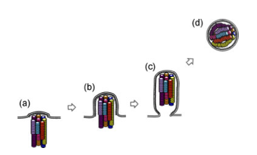
A fancy cartoon showing the exiting virus particle, with bundled genome segments. (Noda et al 2010)

Each one of these studies has potential problems that make them, on an individual basis, insufficient to nail the coffin shut on the random incorporation model. For example, the 7+1 arrangement is only seen in a small fraction of virions. Is it because only some have that arrangement or is it that the preparation of the samples for electron microscopy distorts some particles? This question of how influenza packages its genome serves as a terrific example of the scientific process and the necessity to generate a large body of work to figure out what may seem like a simple question. In class, we talk about designing experiments to test a hypothesis, but tend to see examples of one experiment that really nailed it. It doesn’t usually work that way. You typically need many different experiments, approaching the problem in different ways, because any single experiment could have a valid critique that weakens the conclusion. The whole body of work must be considered, and in this case the body of work appears to be supporting the selective incorporation model.

Share

An epidemic of fear

How does fear of epidemic disease influence the spread of disease?  In the movie “Contagion” we see many examples of how individuals and governments respond to an epidemic.  There is panic, people fleeing cities, people isolating themselves in their homes, governments closing borders and imposing quarantines.   The tagline of the movie, “nothing spreads like fear” may actually be quite accurate.

Using mathematical models to understand the spread of disease is a common tool in epidimiology.  At its most basic, we can think about the spread of a disease through a population by considering that an individual can be susceptible to the disease, infected, or  removed.  Someone who is susceptible can become infected.  Someone who is infected can spread the disease to others who are susceptible.  Infected people can be “removed” from the population by either recovering and becoming immune, or dying.  Either way, they are no longer in the susceptible or infected categories.   These S-I-R models can become quite complicated when you begin to consider all the possible variations:  how easily the disease is transmitted, how long someone is contagious, whether transmission is dependent on direct contact, etc.  These models don’t have to be limited to study the spread of infectious disease: indeed, one could consider fear to be contagious.

So what happens to the spread of disease during an epidemic if we add normal human behavior into the SIR model? If we consider fear to be contagious as well, during an epidemic there there would be two things spreading through the population: fear and disease.  And when we fear disease we change our behaviour.  In a recent paper, the simultaneous spread of fear and disease were modeled using an SIR model.  It turns out that, as the tagline for “Contagion” suggests, fear spreads more quickly than disease.  In their model, someone susceptible to fear can become “infected” with fear either through contact with someone with the disease, someone who has fear but no disease, or someone who has both fear and disease.  Since you can only get infected with the disease by contact with someone else who has the disease, there are more ways to catch fear than disease, and it spreads faster.

So once someone is infected with fear, what do they do?  What would you do? My instinct would be to hide at home and remain isolated; others I have spoken to would flee to a small town in the middle of nowhere.   The researchers considered that too.  How will the spread of the disease change if some people run and some hide and others don’t do anything?  It turns out it is best to hide.  Hiding has the greatest impact on halting the spread of the disease: not only do fewer people become infected, but the disease doesn’t spread as far geographically.  Fleeing reduces the number of people infected (compared to not doing anything) but spreads the disease further.

The model demonstrated another important point.  Over time, our fear decays.  For whatever reasons, after hiding for a while, we may feel more confident about wandering out into the now hopefully less plague ridden world.  Bad idea.  As long as there are still some people infected with the actual disease, re-entering the world adds fuel to the epidemic by introducing more susceptibles.  This causes a second wave of the epidemic.  One could speculate that this could have been the cause of the two waves of influenza in 1918 – people came out of quarantine too early.

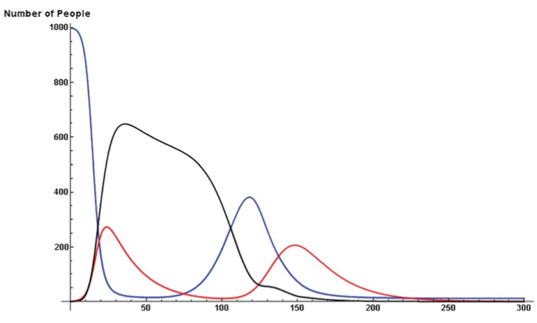
As the number of infected people increase (red), more susceptible people (blue) will hide (black). That causes less people to become infected, people become less afraid and leave quarantine re-entering the population and causing a second wave of disease. From Figure 3 of Epstein et la 2008.

We had a terrific discussion about this paper in class and we identified several reasons why we though that fear would actually spread even faster than the paper suggested, and that the effects of fear (fleeing or hiding) on spread of the disease might be greater.  Obviously, fear can spread without direct contact.  You can get infected with fear pretty easily by reading things on the internet or watching the news.  Fear of a disease could leap across the globe quickly, well ahead of the disease itself.  Also, people don’t behave independently.   So if one member of a family becomes afraid and wants to flee or hide, the rest of the family may do so as well.  As people flee from cities, its likely that choke-points and crowds would further fuel the epidemic.

So in the event of a massive, lethal epidemic, would the politicians and army have the willpower to impose a long enough population-wide quarantine and prevent people from fleeing an infected city?  Would President Obama/Romney say “No really, everyone must stay at home, see there’s this paper in PLoS ONE by Epstein et al…”

Reference:

Epstein, Parker, Cummings and Hammond. (2008). Coupled Contagion Dynamics of Fear and Disease: Mathematical and Computational Explorations.  PLos ONE, 3(12): e3955

Share

Fever!

Most viral infections start out with the same general symptoms: fever, malaise, aches. Those are usually the sign of your immune system starting to fight back. Fever is one of our defense mechanisms, and while it can be quite uncomfortable it is very rarely dangerous. One of the common questions I get asked in my classes is whether it is good or bad to take fever reducing medicines when you are sick. Well, Im not that kind of doctor so I can’t really answer that question, but I can tell you what research has been done on the benefits of fever.

The adaptive value of Saturday Night Fever, caused by listening to Disco, remains unknown

Presumably, by raising your body temperature, you can do a better job of fighting off an infection. The higher temperature may make it more difficult for the pathogen to grow because it grows optimally only at normal body temperature, or it may help the immune system work faster (or both). Some studies in animals have shown that reducing fever results in increased growth of bacteria or virus in the infected animal, and other studies have found increased proliferation, migration and activity of immune cells.

Whatever the mechanism, it is clear that fever must provide some advantage. There are many studies that demonstrate that fever is beneficial in overcoming infection. None of these studies alone is definitive, however taken together, they do seem to support a role for fever in fighting infection. For one, the febrile response is highly conserved in vertebrates (even “cold blooded” animals) and many invertebrates. Some lizards, for example, will seek warmer spots to rest and as a result, raise their body temperature when infected. Fever is also energetically very expensive, requiring 20% more energy to maintain a temperature even a few degrees above normal. It would be unlikely for such an expensive mechanism to be maintained by natural selection if it didn’t have some benefit.

In addition to the evolutionary perspective, several studies in animals show that a fever of a few degrees correlates with better survival rate from infection. Being correlations, we must be cautious in over-interpreting this. Another good way to test if something like fever is useful is to get rid of it. Infected animals can be treated with anti-pyretic (fever reducing) drugs to see what happens to their recovery in the absence of fever. These studies typically show that animals treated with anti-pyretic drugs take longer to recover, and in some cases even to increased mortality. There are some problems with anti-pyretic studies however, and one of the major problems is that many anti-pyretic drugs don’t just reduce fevers. They can have other effects on the body, not all of which are known, and so we can’t always be certain that fever reduction is the reason for the changes in morbidity and mortality.

Of course, I also like to look at the pathogens themselves for hints of what our immune system is doing. They are pretty good at defending themselves against our immune response, so if we look at their defenses we can learn more about how we attack them. The poxvirus Vaccinia encodes a protein that blocks fever. This protein interferes with the function of interleukin-1B, a component of our own immune system that regulates fever. Animals infected with Vaccinia lacking that protein develop fever, showing that when the viral protein is present, the virus can prevent fever. However, interleukin-1B does many other things too, not just regulation of fever, so it is possible that the virus is blocking interleukin-1B for a different reason.

So it is highly likely that fever is good for fighting off infections, but this is not to be taken as medical advice to avoid fever reducing medicines. In the case of naturally occurring infections in humans, we need much more research into fevers resulting from specific infections to decide when a fever is beneficial and should be left alone, whether the fever is dangerous, or if the fever is helpful but the risks of taking an anti-pyretic are worth alleviating the uncomfortable symptoms.

References:
Kluger, M.J. (1996) The adaptive value of fever. Infectious disease clinics of north america. 1(10):1-20.
Alcami, A. and Smith, G.L. (1996) A mechanism for the inhibition of fever by a virus. Proc Natl Acad Sci. 93:11029-11034.

Share

It’s a slime mold, not a cupcake

Slime molds look kind of like vomit but they are pretty awesome.  In fact, one of them is affectionately nicknamed “dog vomit slime mold.”  While vacationing in British Columbia, my 2 year old son, exploring in the trees and shrubs, exclaimed “cupcake!” Knowing it was highly unlikely that there would be a cupcake there, and not wanting him to eat whatever he found, I rushed over to see a pine cone covered in a thick layer of whitish yellowish stuff.  It did, in fact look a little like a piece of rather unappealing cake with thinly smeared white frosting and  sprinkles of pine needles and soil from the forest floor.

Looking around, I found several other similar things growing, and some that looked quite different.  One looked like coral, another had bubbles of clear, sticky liquid.  As  I observed them over several days, they changed, all eventually looking alike, and after more days, dried up completely to a pile of dusty powder that easily fell apart when disturbed.  These were all the same slime mold, at differnt stages of its lifecycle. I’ve sent these photos to various slime mold experts, and it has been tentatively identified as Brefeldia maxima, the “tapioca slime mold.”

Slime molds are indeed slimy, in the picture at the left you can see the slime trail it leaves behind as it moves, but they are not mold.  They are actually amoeba.  These amoeba can exist in the single celled form but under the right conditions, the single cells will amass together. In some slime mold species, called plasmodial slime molds, the cells will actually fuse together to form a single, giant multinucleated cell. Others, called cellular slime molds, form a multicellular mass. Eventually, the slime mold will differntiate and form fruiting bodies, complex strructures in which spores will develop. Spores can then be released to disseminate the slime mold to new locations. What amazes me is the complexity of this process and the blurring of the lines between unicellular and multicellular organisms. You have a single celled organism, in which many individuals can come together into a multicellular (or single giant cell) form, now behaving as one organism that can differntiate to form complex structures like fruiting bodies and spores. Clearly, we can’t be thinking of microorganisms like these as “simple.”

Some recent studies further demonstrate the complexity of slime molds. The slime mold Physarum was used to map an optimal network between points, in this case the Tokyo metropolitan area. Food sources were laid out corresponding to the communities surrounding Tokyo, and Physarum was placed at Tokyo. The slime mold extended plasmodia, or branches, out to the food sources. At first the branches fan out randomly, making many connections, but eventually most plasmodia disappear, leaving only the most optimal connections between the food sources. The final map of the plasmodia turns out to be quite similar to the Tokyo area metro system. Physarum can also do this with the US interstate highway system, and the Canadian highway network (um, isnt there just one highway in Canada?) So an amoeba can simulate a networks developed by human engineers!

In my lab, a student is exploring using Physarum for a similar purpose. Vassar is looking ahead 50 years or so to plan development of the campus. Can we use the slime mold to provide suggestions to the landscape architects on where to place the pathways between buildings? Can the slime mold help me find the best way across campus? We will keep you posted on what we find!

Share

Giant dsDNA Virus Origins: Megaviridae Evolutionary Analysis

Contributed by guest blogger: Katy Hwang ’12

The discovery of the double stranded DNA (dsDNA) virus, Mimivirus, and the subsequent discovery of related Megavirus confounded the size limits of viral particles and the complexity of viral genomes. They are larger or just as large as some bacteria. Megavirus has a 1,259,197-bp genome.  Megavirus contains a genome 6.5% larger than that of Mimivirus. Each of these viruses, classified as Megaviridae, have approximately 979+ proteins, including the first aminoacyl tRNA synthetases (AARS), enzymes that promote translation, found outside of cellular organisms.

Mimivirus infects amoeba; Megavirus natural host is still unknown as it was found in a sea water sample through a campaign on random aquatic environmental sampling off of the coast of Las Cruces, Chile. Megavirus research is conducted in A. castellanii. The virus even reopened the debate on whether or not viruses are alive as Megaviridae have traits that overlap with unicellular organisms such as parasitic bacteria. Phylogenic evidence does not cluster either virus into a prokaryotic clade, but more deeply into a eukaryotic clade. Megavirus is of the archeal type, so it branched out before the radiation of eukaryotes. This provides some insight into the Megaviridae ancestor.  So now the question is: from what did these giant viruses originate and  how did they evolve?

Megavirus and Mimivirus are similar enough to have unambiguous homologous features, but have also diverged enough on the evolutionarily tree to provide more information on the features of a common ancestor. 23% of the Megavirus genome does not have a counterpart in Mimivirus, but it shares about 77% of its 1120 putative coding sequences with Mimivius. Megavirus and Mimivrius use the same motif to specify early gene expression, the expression pattern of the orthologous genes is conserved globally. Not only do these giant viruses have their own AARS, but they also code for their own DNA repair enzymes that correct damage caused by UV light, ionizing radiation and chemical mutagens. Megavirus also has a DNA photolyase, which is an enzyme that uses light energy to make repairs to DNA and increases resistance to UV radiation.

The author’s hypothesis is that the last Megaviridae common ancestor originated from a cellular organism, where the now current Megaviridae have undergone specific genome reduction events. Mimivirus codes for four AARS and Megavirus codes for seven, four being orthologs to those found in Mimivirus and one of the other AARS that Megavirus codes for is a class-II AARS. This observation that viral AARS are not limited to type-I suggests that independent acquisition of these genes by horizontal gene transfer is unlikely.  The ancestor of Megaviridae and Mimivirus most likely started with 20 AARS from a cellular ancestor. It is very unlikely that these seven coding sequences were added, and more likely that there were various lineage specific gene family deletions.

The discovery of the AARS substantiate the claims that the Megaviridae ancestor originated from a cellular organism.  What were the driving factors of these genome reduction events?  More etiological data is required for further analysis of the evolutionary process of the development into these giant dsDNA viruses. What other giant viruses are still out in the world waiting to be discovered and what can we learn from them?

Link:

http://www.pnas.org/content/early/2011/10/04/1110889108


Katy Hwang is a senior at Vassar College, majoring in biology.

Share

H5N1 Ferret Transmission Experiment Published

At last, one of the papers investigating H5N1 influenza transmission in ferrets has been published in the journal Nature yesterday.  To recap the controversy briefly:  news of experimental studies investigating transmissibility of avian H5N1 influenza hit the news this fall, igniting a fierce debate about biosecurity, “dual use” research, and the damage that censorship can have on scientific advancement.  An advisory group, the NSABB, recommended partial censorship of the data, perhaps believing that redacting specific data from the publications would prevent information on how to generate a highly pathogenic mammalian transmissible virus from getting into the hands of bioterrorists or others incapable of handling such viruses safely.  However, after some new data or clarification of data presented in a revised version of the submitted manuscript, the NSABB recommended publication in full.

Avian H5N1 influenza virus has caused sporadic infections in humans who have close contact with infected animals.  Human-to-human transmission has not been observed.  But could an H5 virus mutate or reassort, allowing human-to-human transmission?

One thing that has been lost in this whole controversy is that this study is actually a great demonstration and application of evolution.  How does a virus switch to a new host and transmit efficiently between individuals?  Start with a diverse population that varies in a particular trait (in this case, ability to bind the human receptor).  Put it through selective pressure. This occurred in several steps: first, select for viruses that can bind the human receptor in vitro.  From those that bind, select ones that can efficiently replicate in the respiratory tract of the animal.  Finally, take the efficient replicators and allow for transmission.  At each step of selection, mutations that naturally occur during viral replication further diversify the population resulting in variants that possess the desired property.  Those variants get selected for the next experiment.

This study focused on one particular influenza virus protein, hemagglutinin (HA).  HA is on the surface of the virion and is what the virus uses to attach to the host cell, the first step in viral infection.  HA of human influenza viruses bind a sugar on the surface of human cells, which is slightly different from that found in the avian respiratory tract.  Avian viruses, of course, bind to the form found in birds.  H5 shows a strong preference for binding the avian receptor, so Karaoka et al were interested in finding out if H5 could change to recognize the human receptor, allowing more efficient transmission between mammals.

To address this, they began with a mutagenesis technique to introduce random mutations in the globular head (the receptor binding part) of HA, then used an in vitro approach to select for mutants that bound the human receptor (selection step 1).  Through this process they identified three H5 variants that gained the ability to bind the human receptor while maintaining the ability to bind the avian receptor, and one that switched specificity completely to the human receptor.  Several of the mutations identified in this study had already been shown in previous studies to be important in receptor specificity.

To test if the variant H5s conferred binding to human receptors in vivo, sections of human tracheal tissue were exposed to the viruses and only two were able to infect (selection step 2).  This suggests the virus can infect human epithelium of the upper respiratory tract.

Next, the two remaining variants were used to infect ferrets.  Both replicated in ferret respiratory tracts, but one replicated to higher levels.  When they sequenced the virus that they isolated from that ferret, it was different from what they had put in: a new mutation had appeared.  This new mutation presumably confers the property of better replication in the ferret respiratory tract, so it outgrew the original input virus (selection step 3).

Using this new virus (now with a total of 3 mutations in H5), transmission was tested in ferrets.  Compared to the original H5, which did not transmit via aerosol, the 3-mutation variant did transmit, although between only 2 of 6 animal pairs.  Again, they sequenced the virus that was present in the contact animals and found that it was different than what had gone in to the inoculated animals.  Yet another mutation had appeared (selection step 4).  This additional mutation appears to enhance transmission:  the new virus, now with 4 mutations, transmitted more quickly and between more pairs of animals than the 3-mutation virus.  Although the virus can transmit, none of the infected animals died, but they did show pathology at the site of infection.

So what does this all mean? The best model available for influenza transmission studies is ferrets.  Ferrets aren’t humans, so its important to keep in mind that this is a model that helps us understand what viral or host factors are involved in aerosol transmission in these mammals, and maybe, but not necessarily, in humans.  Since we don’t know what is necessary for human-to-human transmission, it is valuable to have an animal model to give us some ideas of what to look for.  It can provide some good hypotheses on what mediates transmission in humans, which would then have to be further tested.  Obviously, specificity for the human receptor is necessary, but the mutations identified tell us more than that.  Mutations that change specificity are not sufficient for transmission.  It turns out that those mutations also decrease the stability of the HA protein.  The additional mutations acquired through the selection steps compensate for that, and enhance stability.   So now we know that HA stability is important in influenza transmission.  Between ferrets.  That’s probably true for humans too, but it would need to be tested.

If you are still reading, you are obviously procrastinating, and are probably avoiding studying for your final exam.  But here are some more thoughts on the controversy overall.  This is a really interesting paper, with nothing particularly frightening or worrisome about it.  Certainly not any more so than other papers doing similar work that were published without so much controversy.  If “dual use” research needs to be regulated, it needs to be done before the work is done, not after.  If the NSABB was concerned about this kind of research, why only express concern once the experiment succeeds?  In my intro biology class, we read another paper, published in 2005, which was addressing the exact same question, in an almost identical way.  The difference is that they failed to make a transmissible virus.  If there is a concern about this kind of research, a concern that it is too risky to do these kinds of experiments, shouldn’t the alarm have been raised regardless of the outcome?  It just doesn’t make sense to me why it suddenly became so concerning.  If anything good has come of this controversy, it is the widespread discussion that this has stimulated on the importance of open communication of scientific data, the importance of not censoring in science.  Ironically, had we all been given access to the data, like through a journal publication, it would have been apparent that there wasn’t anything to be concerned about.
Share

Teaching and Research on the Microbial World in the Liberal Arts

Social Widgets powered by AB-WebLog.com.