The rise in CO2 levels slows vision and interfers with GABA receptors in damselfish

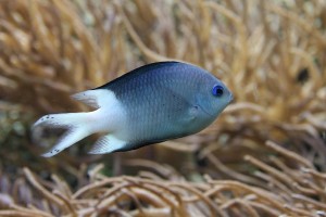
Spiny Damselfish: Acanthochromis polyacanthus

Spiny Damselfish: Acanthochromis polyacanthus

Every day humans are emitting carbon dioxide (CO2) into the atmosphere and causing problems to the environment. The first issue that comes to mind is always global warming and how the earth is going to melt away. I hope you realize by now that this information is false and that there are actual problems that are affecting the way our ecosystem functions.

One actual problem is the rising of carbon dioxide levels in the ocean. This disrupts the marine organisms and may impair the sensory systems and alter the behavior of marine fishes. How can one simple molecule such as CO2 cause all of this?

Molecule of Carbon Dioxide

Molecule of Carbon Dioxide

CO2 is a naturally occurring chemical compound that causes a decrease in the pH of Earth’s oceans, which is known as ocean acidification. When the pH drops, this also affects the fish’s ion balance and disrupts an important neurotransmitter in the brain called GABAA. GABAA is the chief inhibitory neurotransmitter in the central nervous system. Alterations to this inhibitory system can disrupt the visual system of a fish and delay the response time to a predator. This implies that the rise of CO2 levels in the ocean causes impairment in the vision of fish.

Chung et al. (2014) decided to study the affects of CO2 levels on visual responses in damselfish (Acanthochromis polyacanthus) in the Great Barrier Reef. To measure the visual response, Chung focused on vision at the retinal level and how the retina responses to a flickering light.  He measured the critical flicker fusion (CFF) threshold of a fish which is the frequency at which light becomes continuous and the retina stops responding. Typically organisms that are fast moving and live in bright environments have higher CFF  than organisms who spend most of their time in dark environments and are slow.

Chung and his colleagues measured the electrical light response of the retina in damselfish exposed to high levels of CO2 and high levels of CO2. The fish exposed to normal CO2 levels had a high CF of 90 Hz (frequency) while the fish exposed to the high CO2 levels after 6 days had a 68 Hz. This is a great prediction for the future to show if damselfish will have a harder time detecting fast moving objects.

The experiment also tested whether increased CO2 levels in the ocean waters affected GABA signalling system. The GABA receptor was activated by treating the high exposure of carbon dioxide with gabazine As shown in Figure 1 below, the treatment restored the fish’s retinal performance and the CFF threshold increased.

Overall the study showed that when a damselfish is exposed to high levels of CO2, their vision is impacted. However, there are no results indicating what happens in the ecosystem when their vision is impaired. We will need to perform more studies in the future to predict the predation and prey threat in the marine ecosystem

If you want to learn more about the methods and techniques of the experiment you can check out the latest article in the Journal of Experimental Biology.

Chung, W.S., Marshall N. S., Watson, S.A., Munday, P.L., Nilsson G.E. 2014 Ocean acidification slows retinal function in a damselfish through interference with GABAA receptor. J. Exp. Biol. 217: 311-312.

Share
Posted in What's New in Sensory Ecology? | Leave a comment

How do I love thee? Let me smell the ways.

This gallery contains 2 photos.

Love is in the air – quite literally – in the lemur world. A new study has found that the strength of sifaka bonds is reflected by the similarity of their scent signals. Yes, it’s all about chemistry. Researchers from … Continue reading

Share
More Galleries | Leave a comment

The Use of Multisensory Social Information in Birds to Warn Flock Members of Danger

Animals generally live in multisensory worlds, relying upon many different types of information to ensure survival. For animals living in groups who use social information to make decisions, two types of information may be available from other group members: cues and signals. Cues are behaviors inadvertently left by an individual, such as seeing a friend running over to the stove while something is cooking, while signals are behaviors produced by an individual and directed toward others, such as your friend yelling to you that she needs help because the stove is on fire. While both pieces of information are informative, it is hard to say whether they elicit the same response in an individual. Is it possible that having both pieces of information, compared to just one, is advantageous?

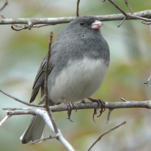
Dark-eyed Junco

Dark-eyed Junco

Randolet et al. (2014) studied the antipredator behavior of the dark-eyed junco, a small American sparrow, which uses alarm calls as an auditory signal and flushing, or fleeing, as a visual cue to warn flock mates of an approaching predator. The researchers predicted that alarm calling and flushing were redundant stimuli, and would therefore elicit the same behavioral response in the juncos. To test this, robotic birds were created to set up an artificial flock, and number of group mates, alarm calling, and/or flushing behavior was manipulated in the robotic birds to resemble varying degrees of risk. Live birds were exposed to these artificial flocks, and their responses to the different levels of risk of the different stimuli were recorded. Change in head position, stretched neck, crouching, movement, flushing, and body turn were all recorded, as these are typical antipredator behaviors of the juncos.

Descriptions of the andipredator behaviors displayed by the juncos.

Descriptions of the andipredator behaviors displayed by the juncos.

The results of the study were contrary to initial predictions, as they showed that multisensory stimuli affect different components of the junco antipredator behavior. In fact, none of the antipredator behaviors performed by the juncos were affected simultaneously by both the alarm calls and flushing behavior, suggesting that these stimuli are non-redundant. Additionally, the degree of alarm of the live birds increased when at least one robot flushed, but reaction times to the robots’ behavior increased, rather than decreased, with at least one alarm call. This suggests possible costs to using each behavior individually, and suggests that, together, these stimuli could facilitate flock cohesion and reduce the occurrence of false alarms to predators.

Although alarm calls and flushing behavior were predicted to be redundant stimuli, it is apparent that there are costs to using each stimulus individually. A flock mate flushing may reflect individuals seeking foraging opportunities, rather than fleeing a predator attack. Thus, if the juncos were to flee the patch each time they witnessed a flock mate flushing, the behavior could waste energy and may result in a loss of foraging opportunities. Additionally, according to the results of the study, alarm calling could actually reduce the benefits of living in a group, by delaying responses to predator attacks. However, it may have been difficult for the researchers in this study to distinguish between alarm calls and low-risk warning calls, which could have caused the results seen.

Randolet et al. (2014) have demonstrated that juncos likely use a non-redundant multisensory system to minimize the occurrence of false alarms using stimuli in different sensory modalities to determine the type of threat. This may ultimately facilitate group cohesion among flocks, allowing for more successful foraging and antipredator behaviors.

Randolet, J., Lucas, J. R., & Fernández‐Juricic, E. (2014). Non‐Redundant Social Information Use in Avian Flocks with Multisensory Stimuli. Ethology

Share
Posted in What's New in Sensory Ecology? | Leave a comment

Oyster Toadfish use stealth signaling to thwart competitors

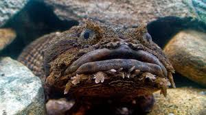
With stiff competition to find that special someone, sometimes it may be necessary to be a little stealthy to beat out your opponents. Male oyster toadfish know this well, according to recent research by Allen F. Mesinger, which examined the use of grunts by male toadfish in order to interrupt the mating calls of competitors.

Toadfish

Many types of animals, such as birds and insects, use acoustic signaling to attract mates. In many of these populations, males adjust their timing so as to not overlap calls, but stealthy toadfish do just the opposite, preferring to interfere with the competitors’ mating calls with their own noises, in a process called “jamming”. The researchers tested the duration, frequencies, and timing of the male toadfish signaling, identifying two separate types, the boatwhistle and the grunt. The boatwhistle consists of a brief grunt, followed by a longer period of pulsing, and is used to attract females. Grunts, however, are shorter, and occur very infrequently spontaneously.

The results showed that 88% of the grunts recorded began and ended during the boatwhistle of another individual, indicating that these grunts were more likely to have been timed according to when the boatwhistles occurred, rather than generated randomly. Additionally, the grunts were consistently emitted during the tonal part of the competitor’s whistle whish has been demonstrated in previous research by Edds-Walton et al. to be the part of the call that attracts females most often. When this part of the call is interrupted, and the frequency digresses from the expected of that range of call, the female finds the call less attractive.

Although it is still unclear whether the grunt serves other purposes, this paper provided strong evidence that an important function is to interfere with the mating attempts of competing males, thus improving the chances of the grunt emitter of finding a mate. This provides a strong example of how sensory (!) cues are important factors in selecting for particular behaviors within a population. 

Mesinger, Allen F. Disruptive communication: stealth communication in the oyster toadfish. The Journal of Experimental Biology 217, 344-350

Share
Posted in What's New in Sensory Ecology? | Leave a comment

Sibling signaling in barn owl nestlings

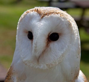
Could we be using bird models to guide some of our own dinner table disputes over who will get the last slice of bread? In a study published this month by the Journal of Evolutionary Biology, researchers from the University of Lausanne in Switzerland suggested that hungry barn owl nestlings (Tyto alba) might be using vocal signals while parents are off hunting, in order to negotiate who will get the next incoming meal.

Barn Owl

Barn Owl

We know that bird songs can help monogamous couples identify their partners and recognize their young and that these songs are often influenced by a combination of genetics and environmental factors, such as learning. But there is still a lot we don’t know about the mechanisms by which individuals recognize and remember calls of their siblings. Previous research has shown that barn owl nestlings have a sibling-sibling communication system that allows them to cooperate in the distribution of food. But how is the fairness of food distribution ensured? Who is keeping track?

The way the sib-sib communication system works is that when the nestlings signal to each other, the nestling that is the most vocal or loudest or vocalizes at the highest frequency is granted priority to the incoming prey without having to compete with it’s siblings. The more food deprived a nestling is, the louder it will compete. The researchers, here, predicted that the signals of barn owl nestlings are individualized and therefore able to be recognized and remembered by their siblings. They suspect that the ability to recognize the individuality of sibling signals might have evolved as a way to strengthen the honesty of the calls. But if the nestlings are all hungry, how is it that the bigger nestlings, which are naturally louder, don’t beat out the smaller ones each time?

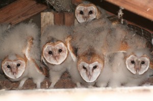
Barn Owl Nestlings

Barn Owl Nestlings

This question gets even more complicated when you take into account the fact that barn owls are asynchronous hatchers: during the breeding season, barn owl moms lay an egg every 2-3 days and the eggs hatch in the order that they are laid so that the oldest and youngest nestlings can be up to a few weeks apart in age. It seems intuitive that the older nestlings, then, would win every vocal competition—but they don’t. In fact, according to what the researchers found, the nestlings actively avoid simultaneous signaling, implying that all siblings want to hear the calls of their nest-mates so that they can recognize their identity and calibrate their own calls in order to compete fairly. The older siblings have been shown to produce fewer negotiation calls when competing with juniors in the nest, seemingly in order to level the playing field. The individual recognition system also ensures that if a nestling cheats, he or she will be punished by the siblings, who will steal the food away.

The bottom line here is that not only do these signals help the nestlings convey their hunger, but the signals have evolved to serve to identify siblings and ensure that the hungriest sibling wins the prize. To learn more, take a look at the original paper in this month’s issue of the Journal of Evolutionary Biology.

Reference: Dreiss, A. N., C.A Ruppli, and A. Roulin. 2014. Individual vocal signatures in barn owl nestlings: does individual recognition have an adaptive role in sibling vocal competition? Journal of Evolutionary Biology 27: 63-75.

Share
Posted in What's New in Sensory Ecology? | Leave a comment

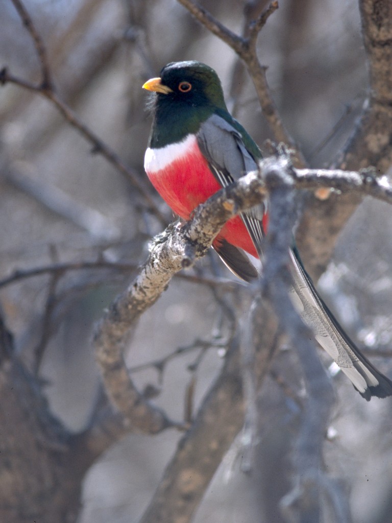
Elegant Trogons: One Gesture for Everyone


Credit: Dominic Sherony

Credit: Dominic Sherony

Imagine coming across a hungry tiger. It fixes its gaze on you, and roars. You have three options: you can stay and fight, you can run, or you can strike your best pose and hope your assailant is deterred. That last option probably wasn’t your first choice; making yourself more enticing is probably the last thing you want.

Visual displays such as posing are common in the animal kingdom. Often, such displays have a target audience. In many species of birds, for example, individuals will threaten each other. Interestingly, such interactions can actually prevent situations from escalating into violence. Visual demonstrations can also be used in attracting and competing for mates. Other displays can deter predators. So, while certain behaviors can be used to signal either within a species (intraspecific) or to another species (interspecific), very few signals straddle that boundary.

But apparently, the elegant trogon (Trogon elegans), a bird native to the southern United States and northern Costa Rica, uses one signal in both courtship and predator-deterrence; it raises its tail to expose the bright red plumage of its underside.

Pierre-Paul Bitton and Stéphanie M. Doucet from the University of Windsor noticed that previous scientific literature had described this tail-raising behavior either in mating or in response to other species. They set out to determine what would prompt this response from elegant trogons, as well as the function of this behavior when used interspecifically.

The Experiments

To get an idea of when elegant trogons raised their tails, Bitton and Doucet spent hundreds of hours in 2010 and 2011 observing them and noting the sex and age of the individuals performing and of conspecifics (members of the same species) in the area. The authors observed that trogons raised their tails to signal each other, both within a sex and to the other sex. They also noted that the birds would raise their tails in response to members of other species, humans, in particular.

Pitton and Doucet also tested the hypothesis that tail-raises could also be used to deter predators by constructing models of the collared forest falcon, a common predator of the trogons. They also constructed models of squirrel cuckoos, a non-threatening bird with a silhouette comparable to that of the falcons, in order to control for variables such as size and shape. The models were placed in strategic locations in order to lure trogons and observe their subsequent responses. They found that trogons were much more likely to raise their tails in encounters with the falcon models than with the non-threatening cuckoo models. Additionally, tail-raises happened much more frequently when the trogons were close to the falcon models.

These two experiments demonstrated that trogons indeed raise their tails to deter predators as well as to communicate with their own species. The evidence supported the conclusion that trogons use the same behavior to attract mates, communicate with each other, and tell predators to back off.

It’s possible that Bitton and Doucet missed something important. Most birds can perceive UV light, which can influence how they appear to other birds based on the UV reflectance or absorbance of their plumage. As the models did not emulate UV reflectance patterns, it’s conceivable that trogons responded differently to the falcon and cuckoo models than they otherwise would have. Given that the trogons responded to a wide range of other potential predators, however, it’s probably a minor oversight.

While there are plenty of examples of multifunctional bird songs that are meant for both intraspecific and interspecific use, this study uncovered a visual signal with the same range of purposes, which is rare. It’s really quite elegant; Need a mate? Dealing with pesky intruders? About to be eaten? Raising your tail is the universal gesture.

Bitton, P. and Doucet, S.M. 2014. A multifunctional visual display in elegant trogons targets conspecifics and heterospecifics. Behavioural Ecology 25: 27-34.

Share
Posted in What's New in Sensory Ecology? | Leave a comment

SAD Ants (Seasonality Affected Decsion-making)

Should I, could I, would I? We make decisions at every moment, every day, every week, every month, and every season. These decisions are influenced by the environment and social interactions. Many animals exhibit social behaviors, such as feeding aggregations … Continue reading

Share
More Galleries | Leave a comment

Insect vision: New insight on a complex circuit

New neurogenic tools of Drosophila research have provided previously unknown information regarding the neural circuitry responsible for visually guided behaviors.  Researchers from the University of Sussex in the United Kingdom have stimulated specific sensory ring neurons using a combined two-photo calcium imaging technique that allowed for in-vivo behavioral observations. Drosophila have compound eyes that have a much lower resolution than camera-like eyes of vertebrates, so how are Drosophila able to extract the right type of information from visual cues?

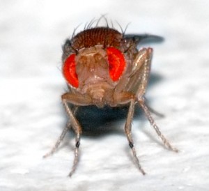
Frontal view of a Drosophila and its compound eyes

Frontal view of a Drosophila and its compound eyes

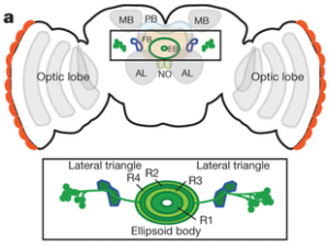
Previous research by Seeling and Jayaraman have proposed a higher brain structure called the central complex which contains a population of visual receptive field neurons. More specifically, they targeted ring neurons R2 and R3/R4d because they receive input from glomeruli. From their experiments, Seeling and Jayaraman were able to target specific cells within the ring neurons that responded to visual stimuli, however they only saw a few of these cells. With only a limited number of cells, it seems that the sparse and coarse encoding capabilities of these cells may not be able to completely reconstruct the visual world. Therefore, the researchers in this article ask what kind of behaviors could be supported by the coarse information encoded by these sensory ring neurons?

Schematic of a Drosophila central brain showing the specific ring neurons (Seeling and Jayaraman, 2013).

Schematic of a Drosophila central brain showing the specific ring neurons (Seeling and Jayaraman, 2013).

The researchers show that R2 cells, when stimulated, show a structured pattern of activation indicating that shape and location are organized within these neurons. Moreover, Drosophila were able to discriminate patterns in an experimental flight simulator. Together with the behavioral assays and the neurogenic tools utilized by Seeling and Jayaraman, the researchers show that R2 neurons are required for the discrimination of at least four visual parameters. Even though the coarse encoding by these cells most likely yields insufficient reconstructions of fine details, enough information is extracted to enable discrimination in the enivornment.

 Researchers have just touched the surface of the complex underpinnings of the sensorimotor behaviors in Drosophila. Further studies examining other sensory processes (such as sound) and their interactions with ring neurons of the central complex may yield even more information regarding Drosophila sensory processes. This article is a great example of how a multifaceted approach including both neurological and behavioral assays can provide value information regarding sensory processing.  For more information on the neurogenic techniques utilized, check out the paper by Seeling and Jayaraman, 2013 in Nature. If you want to learn more about the behavioral assays used in examination of Drosophila visual processing check out the paper in the January 2014 issue of the Journal of Current Biology.

Seelig, J.D., Jayaraman, V. (2013). Feature detection and orientation tuning in the Drosophila central complex. Nature 503:7475, 262-266.

Wystrach, A., Dewar, A.D.M., Graham, P. (2014). Insect vision: Emergence of pattern recognition from coarse encoding. Current Biology 24:2, R78-R80.

Share
Posted in What's New in Sensory Ecology? | Leave a comment

The secret life of the visual cortex: Plasticity in retinal damage and perceptual learning

eyes

Humans are just one of many species that rely heavily on visual information for survival. Accurate vision enables prey and predator detection and recognition of conspecifics, as well as navigational abilities. Because of this reliance on visual cues, damage to the retina (the layer of light-sensitive cells lining the back of the eye) could pose a serious problem. This type of damage can occur as a result of injury, disease, old age, or overexposure to light. So how can animals respond to this impairment to the sensory system?

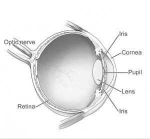
Human_eye_diagram-sagittal_view-NEI

The human eye: The retina lines the back of the eye, and is covered with photoreceptors, which are cells that transmit information about light and color. These cells send this information through the optic nerve to the rest of the brain.

Immediately after the retina is damaged, the region of the visual cortex in the brain that receives information from the injured area stops responding. However, this region slowly recovers activity within a few months. How can this happen if the cells that send information to this region are all dead? Have they come back to life?

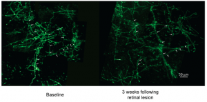
No—the retinal cells themselves cannot recover from damage, but instead, the neurons surrounding the unresponsive brain region can expand and increase the area of the visual field to which they will respond. Although this was already known to occur with excitatory neurons (those that increase neuronal signaling), it was unknown until recently whether inhibitory neurons (those that reduce neuronal signaling) might undergo the same process. By labeling the inhibitory neurons of monkeys (Macaca fascicularis) with a fluorescent tag so that they could be visualized, researchers found that these neurons did indeed undergo immense expansion following damage, in support of their hypothesis (Marik et al., 2014). They observed that after retinal damage had occurred, these normally stable neurons began growing rapidly into the unresponsive region, enabling it to encode visual information from the area of the visual field adjacent to the damage.

Screen shot 2014-02-13 at 11.42.42 PM

Fluorescently-labeled inhibitory neurons in the visual cortex of monkeys. The first image shows the neurons before retinal damage, and the latter shows the same region 3 weeks after the retinal damage. Although the density of the neurons has decreased, the area that they cover has increased (Marik et al., 2014)

These results indicate that inhibitory neurons in the visual cortex are capable of immense structural changes, sometimes expanding to several hundred percent their original size. The authors of this study propose that this expansion of inhibitory neurons could be due to the need to maintain a certain balance between excitatory and inhibitory neurons. Alternately, it could be because these inhibitory neurons are essential for targeting the excitatory neurons that also expand into this region. In any case, because of this process, known as neural plasticity, the part of the brain that used to respond to the damaged region now takes over to enhance the response to the remaining regions of the retina.

Interestingly, researchers further speculate that these mechanisms could be at play during normal sensory experience. In particular, there is some evidence that this type of plasticity is involved in perceptual learning, the process by which organisms can enhance their ability to discriminate between different visual stimuli by repeated exposure, so that very small differences can be detected. This is a crucial aspect of the visual system because it allows organisms to discriminate between various food types (some of which may be lethal) and to recognize mates. This learning mechanism is thought to involve structural changes in the same region of the brain in which the researchers observed neuron growth after retina damage. As a result, many people now believe that the mechanisms involved in neuronal expansion following damage may be the same ones that underlie our ability to train the visual system to make highly specific discriminations that are crucial for survival (Gilbert & Li, 2012).


For more information, see:

Marik, S. A., Yamahachi, H., Borgloh, S. M. A., & Gilbert, C. D. (2014). Large-scale axonal reorganization of inhibitory neurons following retinal lesions. The Journal of Neuroscience, 34(5), 1625-1632.

Gilbert, C. D. & Li, W. (2012). Adult visual cortex plasticity. Neuron, 75(2), 250-264.

Share
Posted in What's New in Sensory Ecology? | Leave a comment

Too fast to see? Touch instead!

If you’re running at 120 body lengths per second, as some species of diurnal tiger beetles (Carabidae: Cicindelinae) can, it would be rather difficult to see everything around you (Figure 1). Motion blur becomes a problem, as it substantially degrades visual contrast and compromises a beetle’s ability to detect obstacles in its path. The fast-running diurnal tiger beetles compensate for this by periodically stopping to reorient itself, especially when hunting down prey. However, entomologists Zurek and Gilbert showed that these insects also perceive the world via antennal touch mechanosensation, which is not affected by motion blur. Moreover, this mechanosensory perception method is both necessary and sufficient for navigation while running.

Figure 1. Hairy-necked tiger beetle, the fast-running diurnal tiger beetle used in Zurek and Gilbert’s study.

Adaptive use of mechanosensory perception in animals with poor vision is common, especially among nocturnal species. Insects living in dark environments use vibrissae and antennae to actively perceive environmental stimuli, including dimensions of a nest site and/or surrounding conspecifics. Diurnal tiger beetles, however, have great daytime vision, but are functionally blind while moving at top speed. Zurek and Gilbert were interested in whether mechanosensory perception can compensate for poor vision due to motion blur, as it does for low light levels, and allow for effective spatial navigation. To answer this question, the researchers first recorded the runs of 20 beetles, each for 10 trials in four different obstacle conditions with varying obstacle heights from 2mm to 4mm; a total of 800 runs. Then, the beetles were separated into four groups, with various physiological impairments (blindness or no antenna) and no impairment. These beetles were subjected to an additional 40 runs in with either obstacles of high contrast or low contrast. Success in obstacle conditions was determined by avoidance of an abrupt collision with the obstacle. Successes with and without antennal contact and failures were averaged in each group (Figure 2).

Figure 2.

Figure 2. Run outcomes, where the control group is the left-most group that is unimpaired and exposed to low-contrast obstacles. High contrast obstacles are marked by a black rectangular box above the beetle. The blinded group is shown with red eyes and the right-most two groups represent the antennectomized groups exposed to different contrast obstacles. Bars represent success/failure rate or runs with and without antenna contact with the obstacle.

Analysis of these runs revealed that presence of antennas and lower obstacle height led to more successful runs, whereas beetle vision or visual contrast of the obstacle had practically no effect on successful runs. Specifically, antennectomized beetles succeeded in as low as 40% of obstacles, while blindness did not affect obstacle success rate. Antennectomized beetles also decreased their ground clearance and pitch angle, suggesting that they may alter their posture to engage antennal contact with the ground in instances when the antenna is short. Such behavior has also been found in cockroaches, which use antennal-ground contact during wall-following. Altogether, the data suggested that the tiger beetles may rely almost exclusively on antennal mechanosensation to avoid collisions while running.

Source:

Zurek, D. B., & Gilbert, C. (2014). Static antennae act as locomotory guides that compensate for visual motion blur in a diurnal, keen-eyed predator. Proceedings of the Royal Society B: Biological Sciences281(1779), doi: 20133072.

Share
Posted in What's New in Sensory Ecology? | Leave a comment