

Humans are just one of many species that rely heavily on visual information for survival. Accurate vision enables prey and predator detection and recognition of conspecifics, as well as navigational abilities. Because of this reliance on visual cues, damage to the retina (the layer of light-sensitive cells lining the back of the eye) could pose a serious problem. This type of damage can occur as a result of injury, disease, old age, or overexposure to light. So how can animals respond to this impairment to the sensory system?
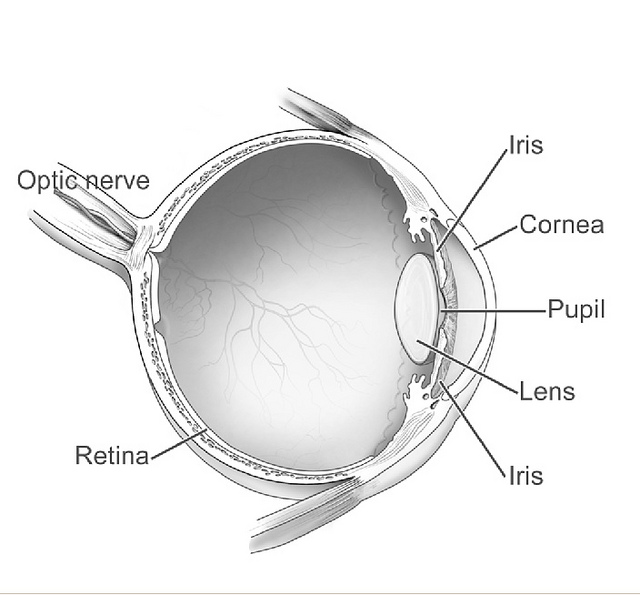
The human eye: The retina lines the back of the eye, and is covered with photoreceptors, which are cells that transmit information about light and color. These cells send this information through the optic nerve to the rest of the brain.
Immediately after the retina is damaged, the region of the visual cortex in the brain that receives information from the injured area stops responding. However, this region slowly recovers activity within a few months. How can this happen if the cells that send information to this region are all dead? Have they come back to life?
No—the retinal cells themselves cannot recover from damage, but instead, the neurons surrounding the unresponsive brain region can expand and increase the area of the visual field to which they will respond. Although this was already known to occur with excitatory neurons (those that increase neuronal signaling), it was unknown until recently whether inhibitory neurons (those that reduce neuronal signaling) might undergo the same process. By labeling the inhibitory neurons of monkeys (Macaca fascicularis) with a fluorescent tag so that they could be visualized, researchers found that these neurons did indeed undergo immense expansion following damage, in support of their hypothesis (Marik et al., 2014). They observed that after retinal damage had occurred, these normally stable neurons began growing rapidly into the unresponsive region, enabling it to encode visual information from the area of the visual field adjacent to the damage.
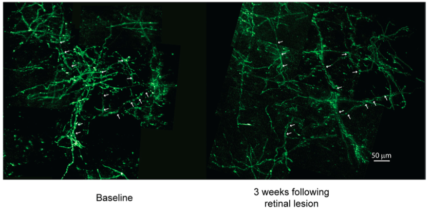
Fluorescently-labeled inhibitory neurons in the visual cortex of monkeys. The first image shows the neurons before retinal damage, and the latter shows the same region 3 weeks after the retinal damage. Although the density of the neurons has decreased, the area that they cover has increased (Marik et al., 2014)
These results indicate that inhibitory neurons in the visual cortex are capable of immense structural changes, sometimes expanding to several hundred percent their original size. The authors of this study propose that this expansion of inhibitory neurons could be due to the need to maintain a certain balance between excitatory and inhibitory neurons. Alternately, it could be because these inhibitory neurons are essential for targeting the excitatory neurons that also expand into this region. In any case, because of this process, known as neural plasticity, the part of the brain that used to respond to the damaged region now takes over to enhance the response to the remaining regions of the retina.
Interestingly, researchers further speculate that these mechanisms could be at play during normal sensory experience. In particular, there is some evidence that this type of plasticity is involved in perceptual learning, the process by which organisms can enhance their ability to discriminate between different visual stimuli by repeated exposure, so that very small differences can be detected. This is a crucial aspect of the visual system because it allows organisms to discriminate between various food types (some of which may be lethal) and to recognize mates. This learning mechanism is thought to involve structural changes in the same region of the brain in which the researchers observed neuron growth after retina damage. As a result, many people now believe that the mechanisms involved in neuronal expansion following damage may be the same ones that underlie our ability to train the visual system to make highly specific discriminations that are crucial for survival (Gilbert & Li, 2012).
For more information, see:
Marik, S. A., Yamahachi, H., Borgloh, S. M. A., & Gilbert, C. D. (2014). Large-scale axonal reorganization of inhibitory neurons following retinal lesions. The Journal of Neuroscience, 34(5), 1625-1632.
Gilbert, C. D. & Li, W. (2012). Adult visual cortex plasticity. Neuron, 75(2), 250-264.
