
New neurogenic tools of Drosophila research have provided previously unknown information regarding the neural circuitry responsible for visually guided behaviors. Researchers from the University of Sussex in the United Kingdom have stimulated specific sensory ring neurons using a combined two-photo calcium imaging technique that allowed for in-vivo behavioral observations. Drosophila have compound eyes that have a much lower resolution than camera-like eyes of vertebrates, so how are Drosophila able to extract the right type of information from visual cues?
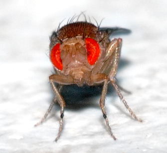
Frontal view of a Drosophila and its compound eyes
Previous research by Seeling and Jayaraman have proposed a higher brain structure called the central complex which contains a population of visual receptive field neurons. More specifically, they targeted ring neurons R2 and R3/R4d because they receive input from glomeruli. From their experiments, Seeling and Jayaraman were able to target specific cells within the ring neurons that responded to visual stimuli, however they only saw a few of these cells. With only a limited number of cells, it seems that the sparse and coarse encoding capabilities of these cells may not be able to completely reconstruct the visual world. Therefore, the researchers in this article ask what kind of behaviors could be supported by the coarse information encoded by these sensory ring neurons?
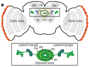
Schematic of a Drosophila central brain showing the specific ring neurons (Seeling and Jayaraman, 2013).
The researchers show that R2 cells, when stimulated, show a structured pattern of activation indicating that shape and location are organized within these neurons. Moreover, Drosophila were able to discriminate patterns in an experimental flight simulator. Together with the behavioral assays and the neurogenic tools utilized by Seeling and Jayaraman, the researchers show that R2 neurons are required for the discrimination of at least four visual parameters. Even though the coarse encoding by these cells most likely yields insufficient reconstructions of fine details, enough information is extracted to enable discrimination in the enivornment.
Researchers have just touched the surface of the complex underpinnings of the sensorimotor behaviors in Drosophila. Further studies examining other sensory processes (such as sound) and their interactions with ring neurons of the central complex may yield even more information regarding Drosophila sensory processes. This article is a great example of how a multifaceted approach including both neurological and behavioral assays can provide value information regarding sensory processing. For more information on the neurogenic techniques utilized, check out the paper by Seeling and Jayaraman, 2013 in Nature. If you want to learn more about the behavioral assays used in examination of Drosophila visual processing check out the paper in the January 2014 issue of the Journal of Current Biology.
Seelig, J.D., Jayaraman, V. (2013). Feature detection and orientation tuning in the Drosophila central complex. Nature 503:7475, 262-266.
Wystrach, A., Dewar, A.D.M., Graham, P. (2014). Insect vision: Emergence of pattern recognition from coarse encoding. Current Biology 24:2, R78-R80.
