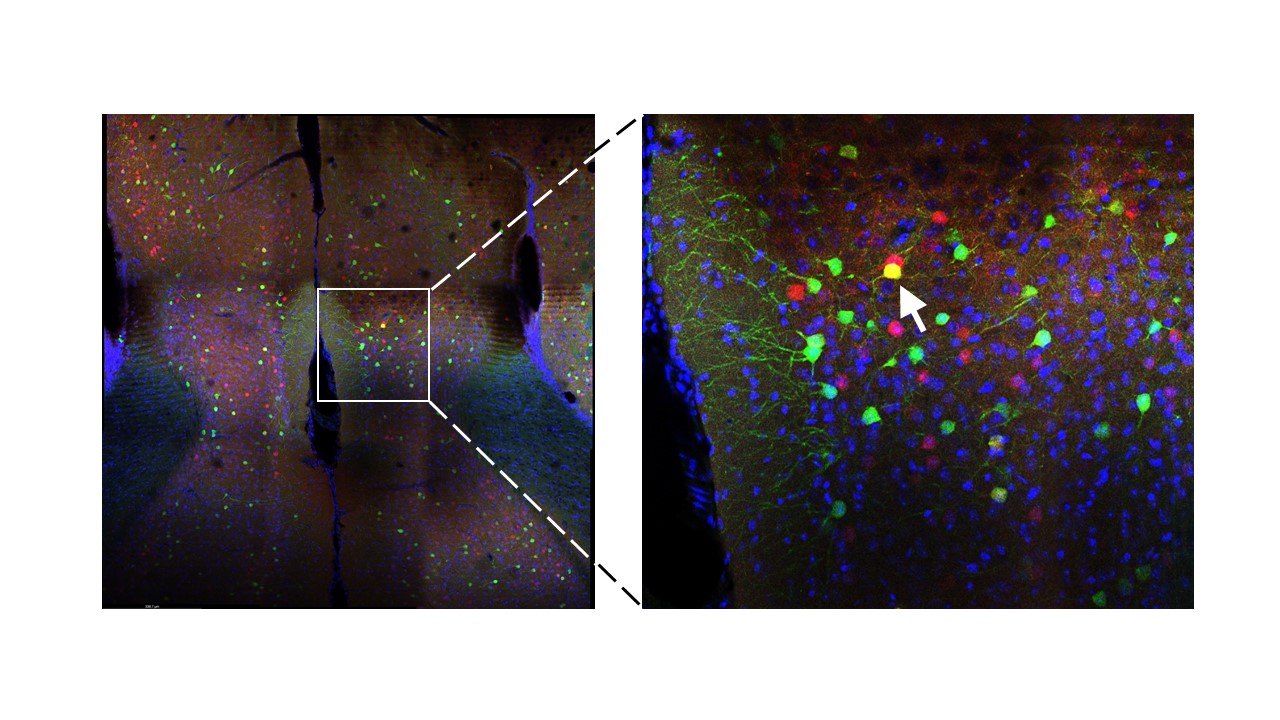
Engram “tagging”
Medial prefrontal cortex tissue immunolabeled for GFP (green), Arc/arg3 (red) and DAPI (blue) using a 20X objective. Co-labeled neuron indicated by arrowhead – the “engram cell”
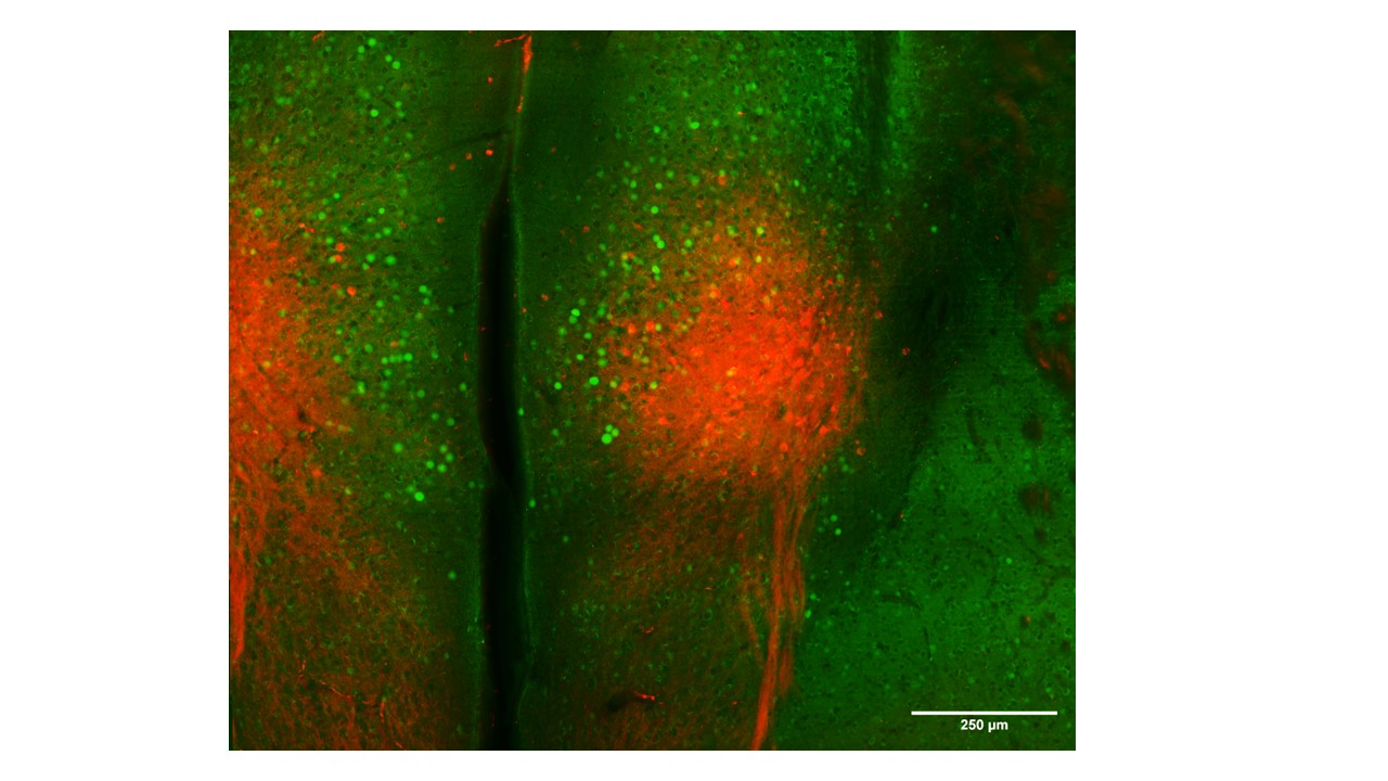
Gi-DREADD (hM4Di) transfection (mCherry) and c-fos staining in the mPFC following fear extinction retrieval.
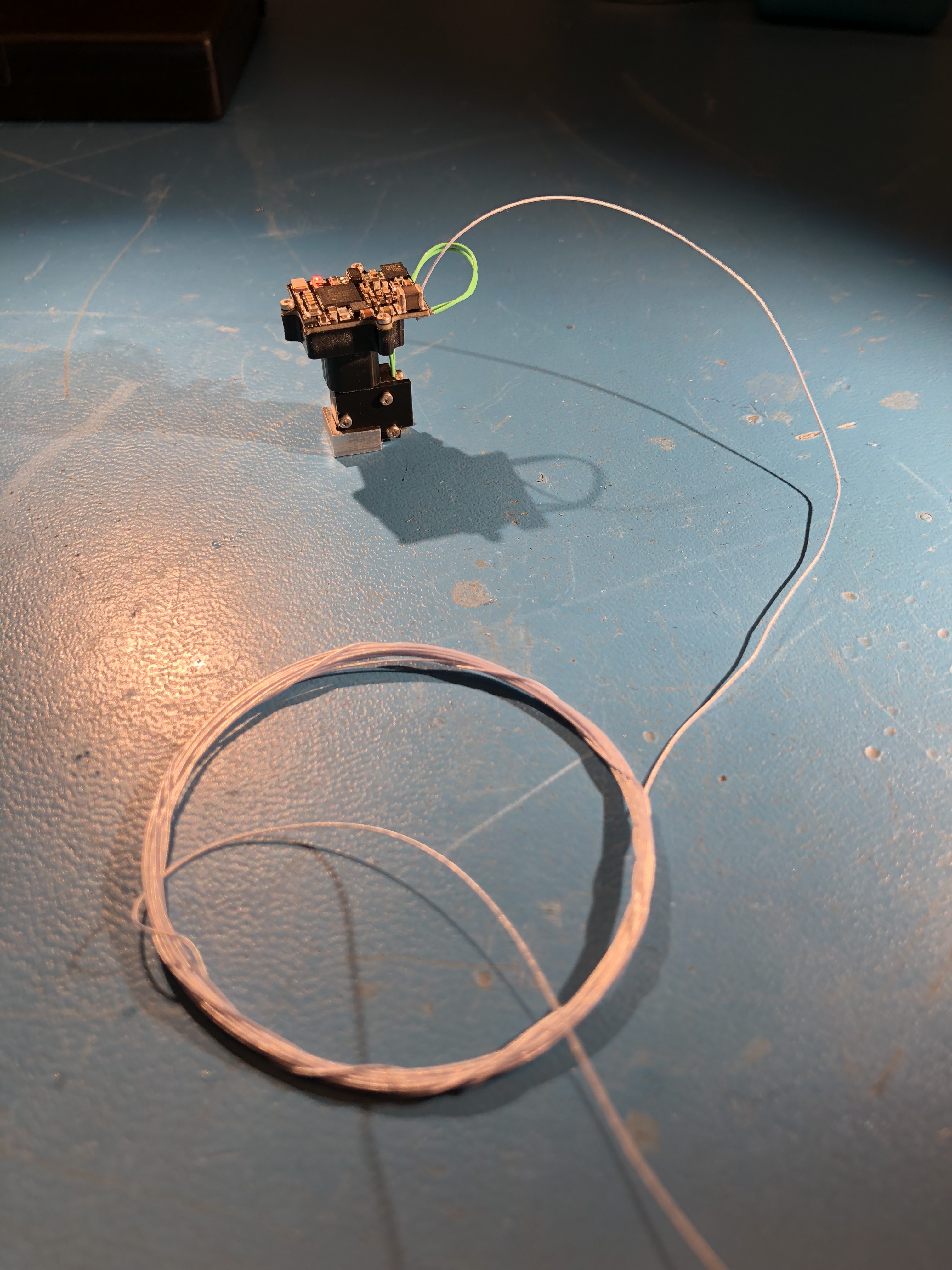
In vivo calcium imaging using miniature microscopes
Infralimbic cortex under conditions that promote behavior extinction.
0.5 mm diameter, 6.1 mm length GRIN. AAV.CamKII.GCamP6s (AAV9).
The miniscope
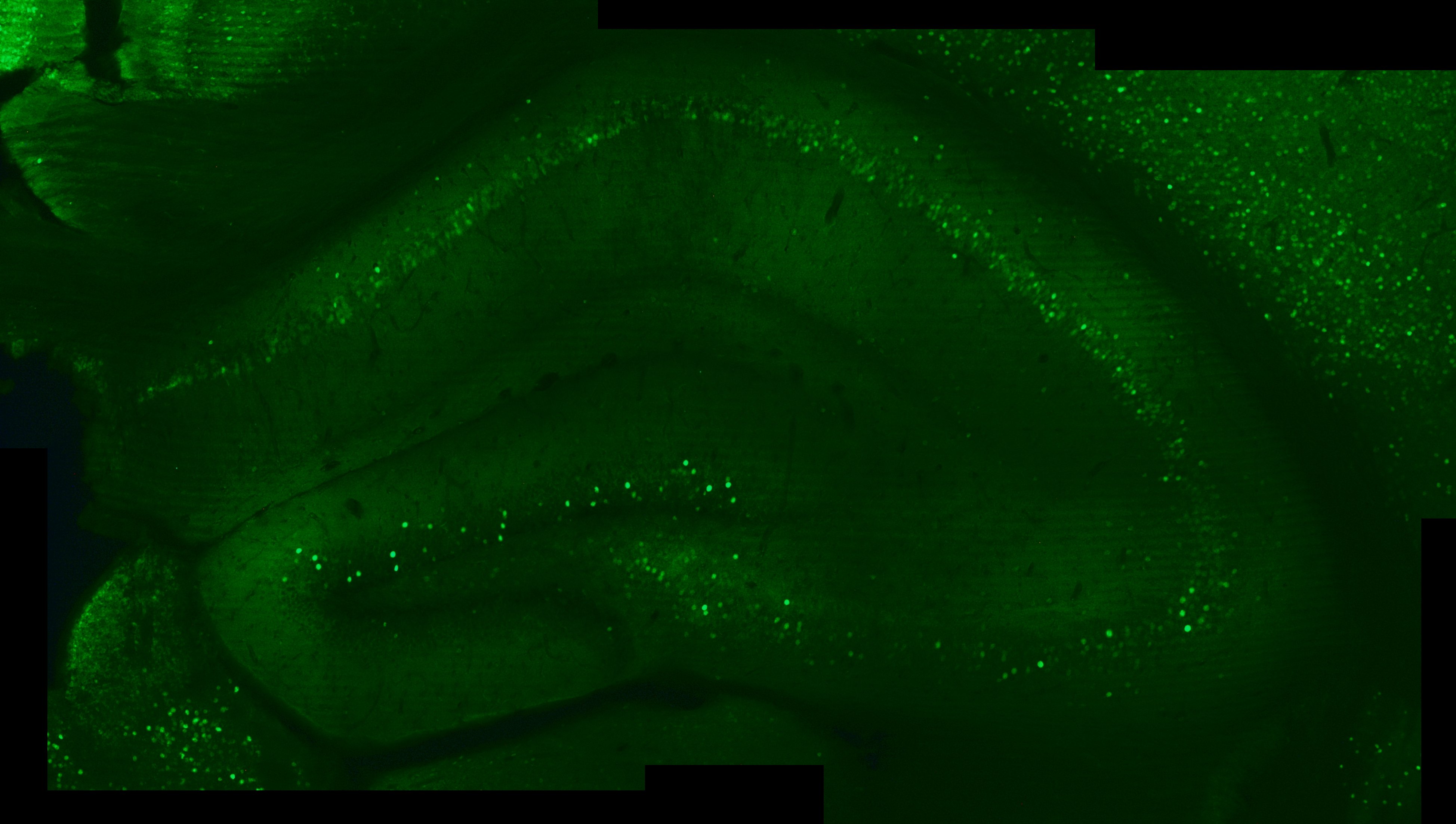
DREADDs (pAAV-CaMKIIa-EGFP)

Golgi-stain
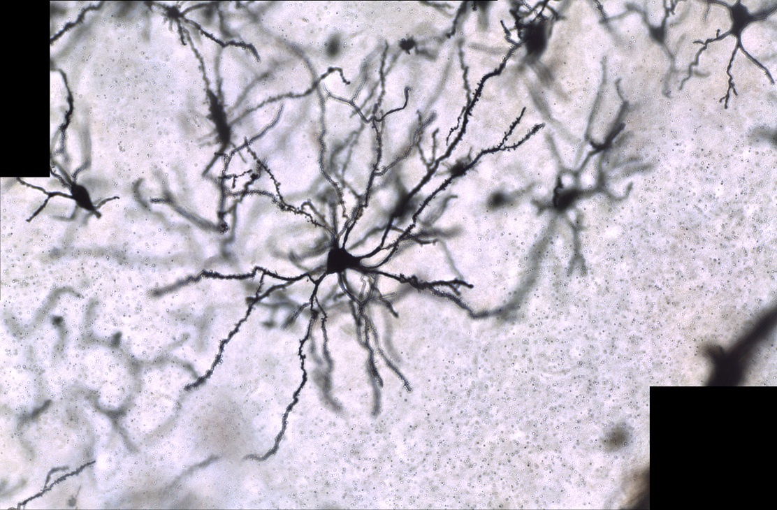
Golgi-stained BLA principal neuron (20 X)
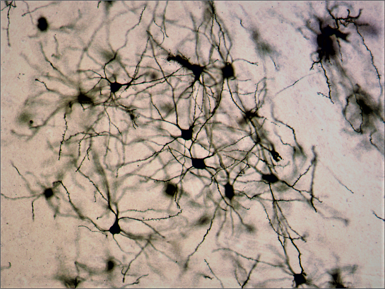
Golgi-stained BLA principal neurons (20 X)
Arc/arg 3.1 immunohistochemistry
c-fos immunofluorescence
3D recontruction of a principal neurons in the basolateral amygdala