Awardees: Mark Schlessman, John Long, Kate Susman
Semester of Award: Spring 2019
Materials Awarded: Canon EF 50mm f/1.8 lens, Canon EOS 6D camera, Smith-Victor copy stand.
Project Description:
What did you hope to learn or achieve with the hardware and/or software you received?
Our goal was to establish common use digital imaging capability for the Department of Biology. We asked for a digital camera, a lens, and a copy stand. We anticipated multiple uses and we cited three in particular:
- Creation of a virtual herbarium (Mark Schlessman)
- Morphometrics of the embryos and egg cases of marine vertebrates (John Long)
- Analysis of nematode locomotion after exposure to pesticides (Kate Susman)
Were you able to put it to the use that you had planned? Please describe.
We did achieve our goal of establishing common use digital imaging capability for our department. The camera and copy stand were set up in part of our common use microscopy area. The equipment is looked after by one of our Science Support Technicians, and all members of the department are aware that it is available to them.
As it has turned out, neither Mark nor Kate has yet used this equipment. Shortly after this grant was funded, Mark acquired an HerbScan (inverted flatbed scanner apparatus) as a gift from the New York Botanical Garden. As it was made specifically for imaging herbarium specimens, the HerbScan is much more convenient than a camera and copy stand. Then, in August 2020, Mark was awarded an NSF grant for herbarium digitization and was able to purchase a camera and light box, which again is more convenient for his purposes than a camera and copy stand. Since Kate became Associate Dean of the Faculty she has had to cut back on research.
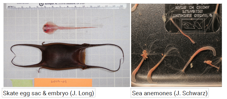
Both John Long and Jodi Schwarz have used the equipment. Here are examples of their images.

What have you learned from this experience that would be useful for other faculty members to know?
Jodi: The camera system is ideal for time lapse photography and video, with very high resolution images. The system is easy to program and intuitive to use. The lighting system allows for variable lighting that can be supplemented with other light sources, such as LED and incandescent light sources. The system is also very sturdy. It is ideal for student projects, due to its ease of use. It is also very easy to transport on a cart to other locations, allowing people to share easily.
John: Flexible and high-resolution imaging — still and video — is useful for a range of projects. The detachable camera adds to flexibility. The diffuse lighting is also helpful for reducing sharp visual gradients.
What impact would you say this grant has had on your students or your research?
This camera system supported two URSI projects and several independent student projects focusing on spawning and reproduction in sea anemones.
John: Imaging studies are a great way to engage students. The training is rapid, the results are immediate, and the skills are transferable. Two students worked on the skate project, and they co-presented their work at a scientific conference: “Behavior of the encapsulated embryos of little skates, Leucoraja erinacea,” co-presented by Connor McShaffrey (VC ’21) and Eden Forbes (VC ’21). Annual meeting, Society for Integrative and Comparative Biology (SICB), remote conference, Jan-Feb 2021.
