This page was written by Rebecca Eells ’12
We are now at Delaware State University working in the optics lab. While there are multiple projects being conducted, one of the main projects is LIBS. LIBS stands for laser-induced breakdown spectroscopy. A highly energetic infrared laser pulse is used to create a plasma on the surface of the sample. As the excited atoms return to ground state characteristic photons are released. The optical fiber detects the photons and allows the AutoLibs program (written in LabView) to detect the atomic spectra. By analyzing the spectra, the individual elements in the sample can be determined.
The first sample that was used in the LIBS experiment was an untreated silicon wafer. This untreated wafer will be used as a control. The next sample that was run was a silicon wafer with quantum dots on the surface (sample J06). Quantum dots are nanoparticles that are part of a special class of semiconductor. These quantum dots emit photons under excitation, which are visible as light to the human eye. The wavelength of the emission photons depends on the size of the quantum dot, not the material.
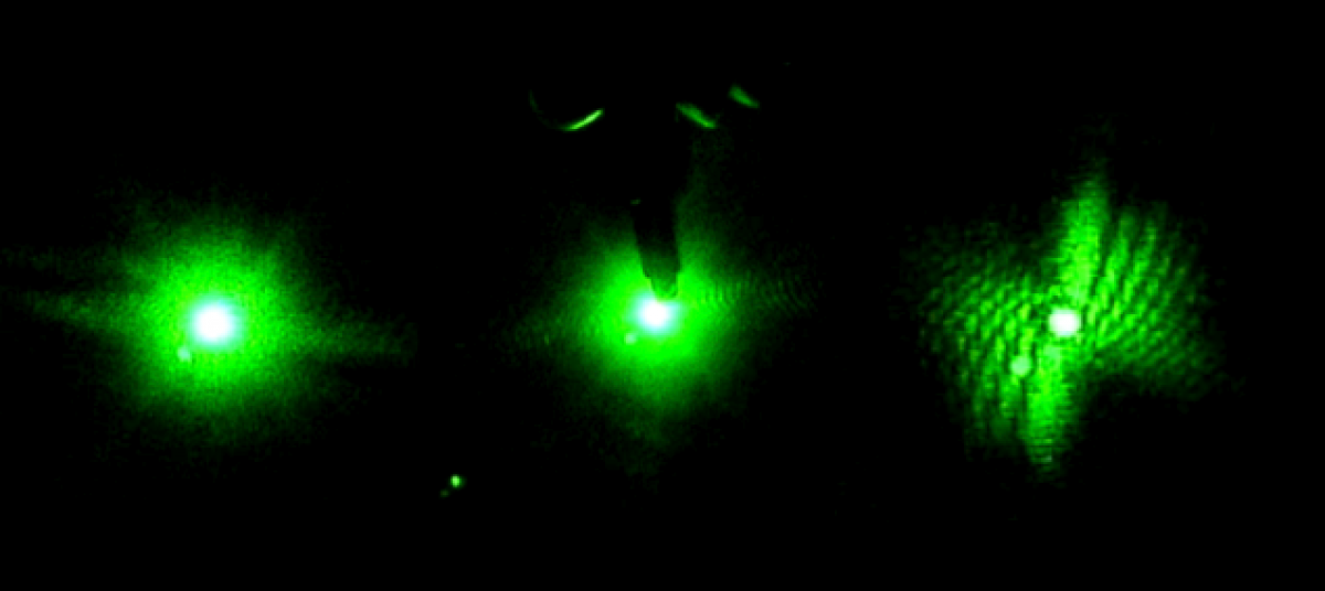
Below is a picture on the J06 sample. The quantum dots are located in a circle right in the middle of the silicon wafer.

All samples were rinsed with distilled water three times in a glass vial. The vial was then filled halfway with distilled water and washed in the ultrasound machine for 10 minutes. Each sample was then taped (using double stick tape) onto a piece of cardstock. The cardstock was then taped to an aluminum cylinder.
The LIBS system was calibrated using a blank piece of cardstock. The optical fiber was adjusted as a scan of the system was being run to maximize the intensities. Once the system was calibrated, the control sample was placed on the stage. The laser was set to two energy units to prevent the sample from being destroyed. The AutoLibs was set to fire 100 shots at the sample. The shots are fired in a spiral as the sample is slowly rotated. The diameter of the shots was set for diameter one, which is the smallest diameter. The control data was saved to the computer and the same process was repeated for the J06 sample.
Once data was obtained for both samples, a graph was made in Origin. The X-axis is wavelength (the same for every sample) and both the control and the J06 sample were represented on the Y-axis.

The data for J06 is now ready to be analyzed and hopefully the elements that are present can be determined.
Thank you to our collaborators:
Dr. Yuri Markushin, Delaware State University
Harry Burton, Undergraduate Student at Delaware State University