
In 1959, Lettvin and colleagues first suggested that the eye does not simply transmit images like a camera; rather, important processing happens at the level of the eye — for example, the extraction of valuable visual information from noise. This became a seminal paper in retinal research and the basis for Baden and colleagues’ new question:
How is this visual information processed and how many different kinds of retinal ganglia cells (RGCs) are involved?
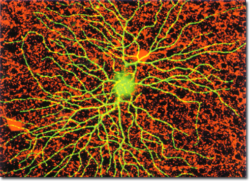
Retinal ganglia cell (usdbiology.com)
Output from the retina is transmitted via many types of RGCs, each unique in the kind of visual features they transmit to the brain. Traditionally they have been classified by whether they are stimulated by brightening (“ON”) or dimming (“OFF”) of the receptive field. Each brand of cell transmits information via a unique “channel.” However, the exact nature and number of these channels is still misunderstood.
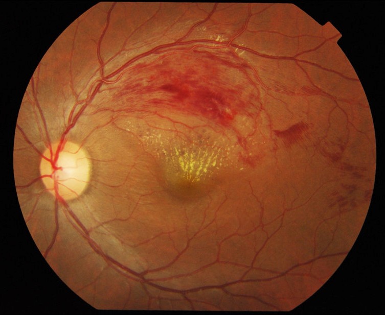
Color fundus photograph of left eye showing retinal vein. (https://commons.wikimedia.org/wiki/Retina#/)
To begin answering this question, Baden and colleagues took on the largest RGC analysis to date, examining over 11,000 mouse retinal cells (compared to the ~450 previously examined) using a combination of electrical stimulation and recording, staining, and dendritic reconstruction. Researchers stimulated each retinal cell with four kinds of light, each targeting either contrast, polarity, direction, color, or other categories of visual information.
They then reconstructed the cells using electroporation, a technique in which an electrical field increases the permeability of the cell membrane allowing substances, in this case a fluorescent stain, to enter each cell. With this technique they were able to determine the size, position, and behavior of each of the targeted cells.

Blue eye of boy ((https://commons.wikimedia.org/wiki/Blue_Eye_Boy/
Baden and colleagues identified approximately 40 different kinds of channels out of the retina. This included all previously discovered channels and approximately 20 new ones – a huge step forward in this line of research. This is particularly significant because the increased variety of channels suggests even more variety in cells themselves. The discovery of increased cellular diversity in the retina brings us one step closer to understanding the ways in which the retina communicates with the brain. It also posits a need for far more classifications within RGCs than previously anticipated. While this paper did not look into a mechanistic analysis of the channels within the retina, it is an important step forward in understanding and categorizing the major players within the visual system — at least in mice. If generalized, this research has the potential to aid in the creation of artificial visual systems or cure visual anomalies at the retinal level.
References
Baden, T., Berens, P., Franke, K., Rosón, M. R., Bethge, M., & Euler, T. (2016). Article The functional diversity of retinal ganglion cells in the mouse. Nature, 529(7586), 345–350.
Lettvin, J. Y., H. R., Maturana, W. S., McCulloch, W. H., Pitts. (1959). What the Frog’s Eye Tells the Frog’s Brain. Proceedings of the IRE, (47), 233-258.
